. 2010 Jul 1;21(13):2182-90.
doi: 10.1091/mbc.e10-02-0154. Epub 2010 Apr 28.

Brain-derived neurotrophic factor regulates satellite cell differentiation and skeltal muscle regeneration

Affiliations

Brain-derived neurotrophic factor regulates satellite cell differentiation and skeltal muscle regeneration

Charlene Clow et al. Mol Biol Cell. .

Abstract

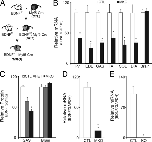
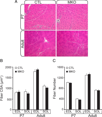
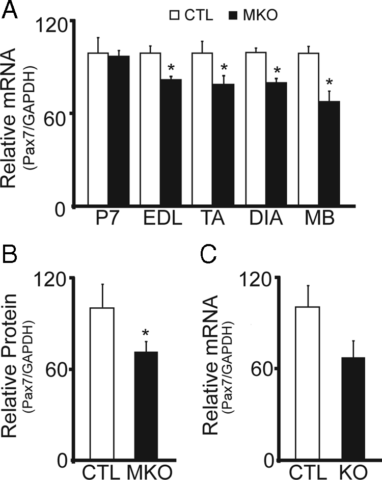
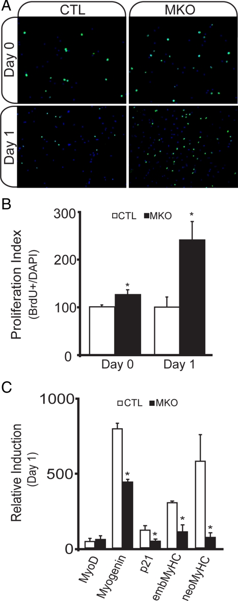
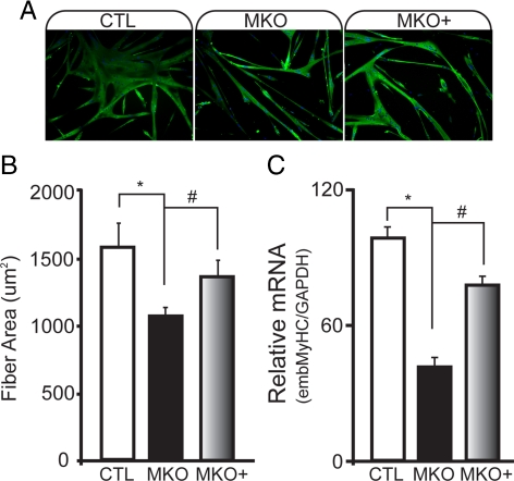
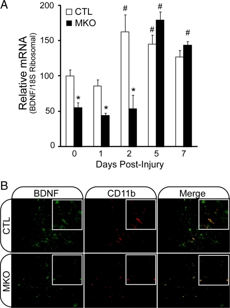
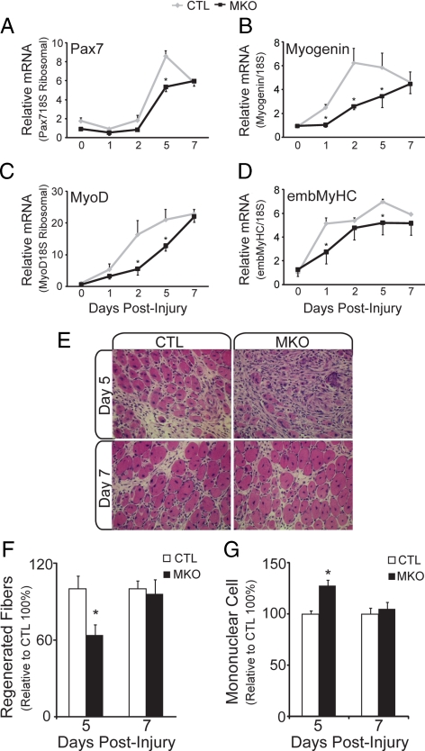
In adult skeletal muscle, brain-derived neurotrophic factor (BDNF) is expressed in myogenic progenitors known as satellite cells. To functionally address the role of BDNF in muscle satellite cells and regeneration in vivo, we generated a mouse in which BDNF is specifically depleted from skeletal muscle cells. For comparative purposes, and to determine the specific role of muscle-derived BDNF, we also examined muscles of the complete BDNF(-/-) mouse. In both models, expression of the satellite cell marker Pax7 was significantly decreased. Furthermore, proliferation and differentiation of primary myoblasts was abnormal, exhibiting delayed induction of several markers of differentiation as well as decreased myotube size. Treatment with exogenous BDNF protein was sufficient to rescue normal gene expression and myotube size. Because satellite cells are responsible for postnatal growth and repair of skeletal muscle, we next examined whether regenerative capacity was compromised. After injury, BDNF-depleted muscle showed delayed expression of several molecular markers of regeneration, as well as delayed appearance of newly regenerated fibers. Recovery of wild-type BDNF levels was sufficient to restore normal regeneration. Together, these findings suggest that BDNF plays an important role in regulating satellite cell function and regeneration in vivo, particularly during early stages.


Figures

Figure 1.
Figure 1.
Generation of the muscle-specific BDNF knockout mouse. (A) Schematic representation of the breeding scheme used in generation of the muscle-specific BDNF knockout mouse. (B) Quantification of the relative levels of BDNF mRNA detected by qRT-PCR in hindlimb musculature at P7 and from adult EDL, GAS, TA, SOL, and DIA muscles of BDNFwt/wt; Myf5-Cre (CTL), and BDNFf/f; Myf5-Cre (MKO) animals (n = 6; *p < 0.01). (C) Relative levels of BDNF protein per milligram wet tissue mass in brain and GAS muscle preparations from adult CTL, HET and MKO animals (n = 3; *p < 0.05). (D) Relative levels of BDNF mRNA detected in CTL and MKO single fiber myoblast preparations cultured for 5 d in growth-promoting medium. (E) Relative levels of BDNF mRNA detected in hindlimb musculature of BDNF−/− (KO) compared with control littermates (CTL) at P1 (n = 3; *p < 0.001). Error bars represent SE.
Figure 2.
Figure 2.
Overall muscle histology is not affected in the absence of muscle-BDNF. (A) Representative hematoxylin- and eosin-stained cross sections from control (CTL) and BDNFMKO (MKO) hindlimb sections at P7 (top) and adult muscle (bottom). (B) Quantification of the average cross-sectional area. (C) Fiber number from P7 and adult EDL and SOL muscles (n = 6). Error bars represent SE.
Figure 3.
Figure 3.
Decreased expression of the satellite cell marker, Pax7, in the absence of muscle-BDNF. (A) Quantification of the relative levels of Pax7 mRNA in P7, adult EDL, TA, and DIA muscles, and single fiber myoblast preparations (MB) from BDNFMKO (MKO) and control littermates (CTL). (B) Quantification of Pax7 protein levels standardized to GAPDH (n = 6; p < 0.05). (C) Relative levels of Pax7 mRNA in BDNF−/− (KO) compared with control littermates (CTL) at P1 (n = 3). Error bars represent SE.
Figure 4.
Figure 4.
Abnormal satellite cell proliferation in single fiber myoblast preparations from BDNFMKO mice. (A) Representative BrdU labeling (green) of proliferating (day 0, top) and early differentiating (day 1, bottom) myoblasts isolated from BDNFMKO (MKO) and control (CTL) littermates. Images are counterstained with 4′,6-diamidino-2-phenylindole (DAPI) in blue. (B) Quantification of the ratio of BrdU-labeled nuclei relative to the total number of nuclei per field of view (n = 6, *p < 0.05). C) Relative induction of MyoD, myogenin, p21, embryonic (emb), and neonatal (neo) MyHC transcripts compared with proliferating cultures after 1-d exposure to differentiation promoting medium (n = 6; *p < 0.05). Error bars represent SE.
Figure 5.
Figure 5.
Decreased size and MyHC expression in BDNF-depleted myotubes is rescued by treatment with exogenous BDNF protein. (A) immunofluorescence labeling of total MyHC in control (CTL), BDNFMKO (MKO), and MKO cultures treated with 20 ng/ml recombinant BDNF protein (MKO+) after 5-d differentiation. Counterstaining with 4′,6-diamidino-2-phenylindole (DAPI) was performed. (B) Quantification of the average area of MyHC-positive myotubes. (C) Relative MyHC mRNA levels standardized to GAPDH after 5-d differentiation (n = 6; *, significant decrease compared with CTL; #, significant increase compared with MKO; p < 0.05). Error bars represent SE.
Figure 6.
Figure 6.
BDNF expression by CD11b-positive cells in regenerating skeletal muscle. (A) Quantification of the relative levels of BDNF transcript in regenerating BDNFMKO (MKO) and control (CTL) muscles at 0, 1, 2, 5, and 7 d postcardiotoxin injection (n = 6; *, decrease and #, increase compared with controls at day 0; p < 0.05). (B) immunofluorescence labeling of BDNF (green) and CD11b (red) in regenerating TA muscles of MKO and CTL animals 5 d after injury.
Figure 7.
Figure 7.
Delayed regeneration in the absence of muscle-BDNF. Quantification of the relative levels of Pax7 (A), myogenin (B), MyoD (C), and embMyHC (D) transcript in regenerating BDNFMKO (MKO) and control (CTL) muscles. All values are relative to control day 0 transcript levels (n = 6; *p < 0.05). (E) Representative hematoxylin and eosin staining of regenerating TA muscles from CTL and MKO animals at days 5 (top) and 7 (bottom) after injury. (F) Quantification of the number of newly regenerated (centrally nucleated) fibers in CTL and MKO muscles at 5 and 7 d after injury. (G) Relative number of mononuclear cells within regenerating MKO and CTL muscles at 5 and 7 d after injury. All values are relative to control levels (n = 3; *p < 0.05). Error bars represent SE.

References

    1. Capsoni S., Ruberti F., Di Daniel E., Cattaneo A. Muscular dystrophy in adult and aged anti-NGF transgenic mice resembles an inclusion body myopathy. J. Neurosci. Res. 2000;59:553–560. - PubMed
    1. Carrasco D. I., English A. W. Neurotrophin 4/5 is required for the normal development of the slow muscle fiber phenotype in the rat soleus. J. Exp. Biol. 2003;206:2191–2200. - PubMed
    1. Charge S. B., Rudnicki M. A. Cellular and molecular regulation of muscle regeneration. Physiol. Rev. 2004;84:209–238. - PubMed
    1. Chevrel G., Hohlfeld R., Sendtner M. The role of neurotrophins in muscle under physiological and pathophysiological conditions. Muscle Nerve. 2006;33:462–476. - PubMed
    1. Condrea E. Membrane-active polypeptides from snake venom: cardiotoxins and haemocytotoxins. Experimentia. 1974;30:121–129. - PubMed

Publication types

MeSH terms

Substances

LinkOut - more resources