. 2010 Jun 15;21(12):1937-44.
doi: 10.1091/mbc.e10-02-0101. Epub 2010 Apr 28.

Ribosome-binding proteins Mdm38 and Mba1 display overlapping functions for regulation of mitochondrial translation

Affiliations

Ribosome-binding proteins Mdm38 and Mba1 display overlapping functions for regulation of mitochondrial translation

Heike Bauerschmitt et al. Mol Biol Cell. .

Abstract

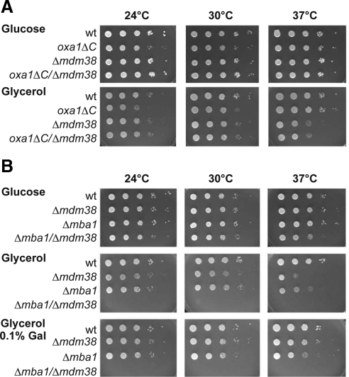
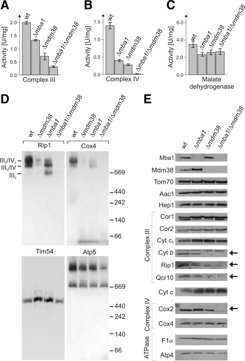
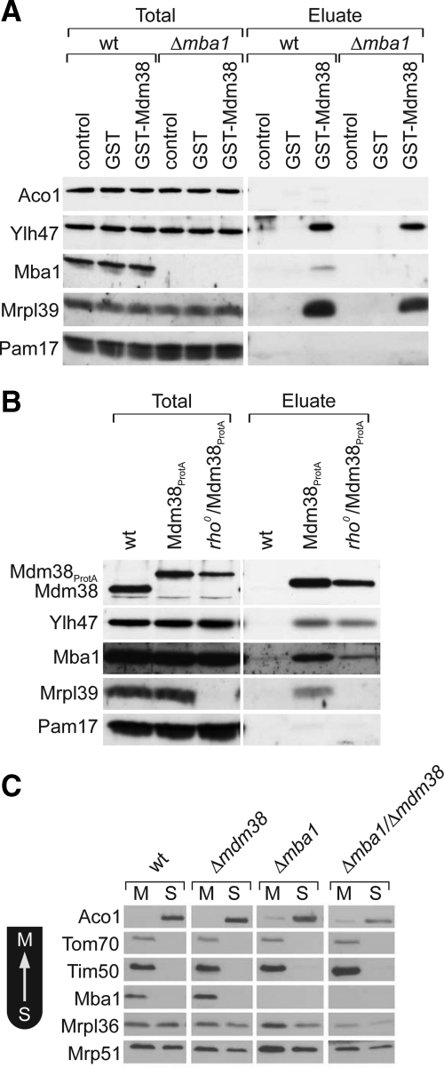
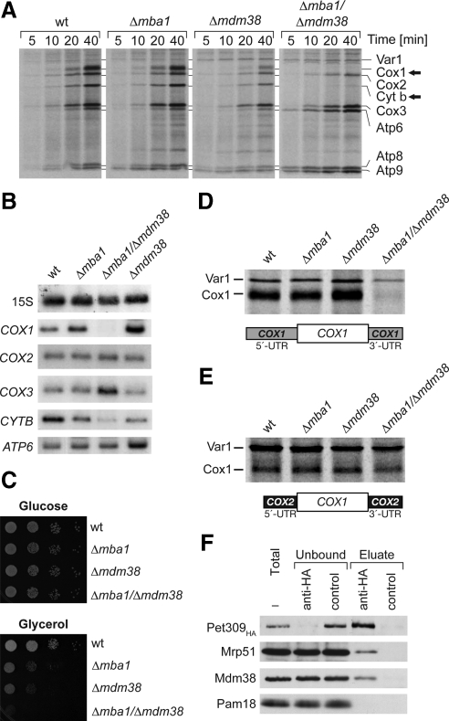
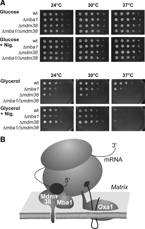
Biogenesis of respiratory chain complexes depends on the expression of mitochondrial-encoded subunits. Their synthesis occurs on membrane-associated ribosomes and is probably coupled to their membrane insertion. Defects in expression of mitochondrial translation products are among the major causes of mitochondrial disorders. Mdm38 is related to Letm1, a protein affected in Wolf-Hirschhorn syndrome patients. Like Mba1 and Oxa1, Mdm38 is an inner membrane protein that interacts with ribosomes and is involved in respiratory chain biogenesis. We find that simultaneous loss of Mba1 and Mdm38 causes severe synthetic defects in the biogenesis of cytochrome reductase and cytochrome oxidase. These defects are not due to a compromised membrane binding of ribosomes but the consequence of a mis-regulation in the synthesis of Cox1 and cytochrome b. Cox1 expression is restored by replacing Cox1-specific regulatory regions in the mRNA. We conclude, that Mdm38 and Mba1 exhibit overlapping regulatory functions in translation of selected mitochondrial mRNAs.


Figures

Figure 1.
Figure 1.
Yeast cells lacking Mba1 and Mdm38 show severe synthetic growth defects. (A and B) Wild-type (wt) and mutant yeast strains were grown to midlog phase, adjusted to an OD600 of 0.1, and 10-fold serial dilutions spotted onto plates containing 2% glucose, 2% glycerol, or 2% glycerol and 0.1% galactose (Gal). Plates were incubated for 2 (glucose) or 5 d at the indicated temperatures.
Figure 2.
Figure 2.
Δmba1mdm38 mitochondria show severe defects in complexes III and IV of the respiratory chain. (A–C). Complex III, complex IV, and malate dehydrogenase activities were measured in isolated mitochondria from the strains indicated. SEs were calculated from three independent experiments. (D) Mitochondrial protein complexes were resolved by BN-PAGE, transferred to PVDF membranes, and probed with antibodies against Rip1 (complex III), Cox4 (complex IV), Tim54 (TIM22 complex), and Atp5 (FoF1-ATPase). Positions of molecular-weight markers in kDa are indicated. (E) Mitochondria (50 μg) of the indicated strains were analyzed by Western blotting with antibodies against the indicated proteins. Cyt b, cytochrome b; Cytc1, cytochrome c1; F1α, α subunit of the FoF1-ATPase. The arrows indicate proteins that show diminished levels in the double mutant.
Figure 3.
Figure 3.
Mba1 and Mdm38 physically interact with each other and with mitochondrial ribosomes. (A) Wild-type (wt) and Δmba1 mitochondria were lysed and incubated with purified GST or GST-Mdm38 bound to glutathione Sepharose. After extensive washing, bound proteins were eluted and visualized by Western blotting using antibodies against the indicated proteins. Control lanes show an aliquot of the mitochondrial extracts. (B) Wild-type or Mdm38ProtA-expressing mitochondria that contain or lack a mitochondrial genome were lysed with buffer containing 1% digitonin. The extracts were incubated with IgG Sepharose. The resin was washed and bound proteins were eluted and analyzed by Western blotting. Four percent of the total sample and 100% of the eluate were loaded on the gels. (C) Mitochondria of the indicated strains were fractionated into membrane (M) and soluble (S) fractions by freeze-thawing and floatation. Proteins of these fractions were analyzed by Western blotting.
Figure 4.
Figure 4.
Δmba1mdm38 mitochondria synthesize severely reduced amounts of Cox1 and Cyt b. (A) Mitochondria isolated from the strains indicated were incubated in [35S]methionine-containing translation buffer for the times indicated. Mitochondria were reisolated, washed, and subjected to SDS-PAGE and autoradiography. Arrows depict Cox1 and Cyt b. (B) Δmba1mdm38 mitochondria lack COX1 mRNA. RNA was isolated from the indicated mitochondria and analyzed by Northern blotting using radioactive probes for the indicated transcripts. (C) Wild-type (wt) and mutant yeast strains lacking mitochondrial introns were grown to midlog phase, and 10-fold serial dilutions were spotted onto plates containing glucose or glycerol/ethanol. Plates were incubated at 30°C. (D and E) Expression of Cox1 under the COX2 promoter bypasses the need for Mba1 and Mdm38. Mitochondrial translation products were radiolabeled in the indicated deletion mutants. Cells contained a wild-type mitochondrial genome (D) or the reading frame of COX1 was flanked by UTRs from COX2 (E). (F) Mdm38 interacts with the Cox1-specific translational activator Pet309. Mitochondria carrying Pet309HA were solubilized in digitonin buffer and subjected to immunoprecipitation with anti-HA or anti-FLAG antibodies (control). Bound protein was analyzed by Western blotting. Five percent of total and unbound samples and 100% of the eluates were loaded on the gels.
Figure 5.
Figure 5.
The Δmba1mdm38 growth defect is independent of a role of Mdm38 in the maintenance of mitochondrial K+/H+ homeostasis. (A) Growth of the indicated strains was analyzed as described for Figure 1A. Plates labeled +Nig contained 2 μM nigericin. (B) Hypothetic model for the role of Mdm38 and Mba1 in translational control. Ribosome-associated membrane proteins are depicted. Translational activators (black sphere) associate specifically with the 5′-UTR of mitochondrial transcripts and might employ Mba1 and Mdm38 for their recruitment to mitochondrial ribosomes.

References

    1. Altmann K., Dürr M., Westermann B. Saccharomyces cerevisiae as a model organism to study mitochondrial biology: general considerations and basic procedures. Methods Mol. Biol. 2007;372:81–90. - PubMed
    1. Barrientos A., Korr D., Tzagoloff A. Shy1p is necessary for full expression of mitochondrial COX1 in the yeast model of Leigh's syndrome. EMBO J. 2002;21:43–52. - PMC - PubMed
    1. Bauerschmitt H., Funes S., Herrmann J. M. The membrane-bound GTPase Guf1 promotes mitochondrial protein synthesis under suboptimal conditions. J. Biol. Chem. 2008;283:17139–17146. - PubMed
    1. Dimmer K. S., Fritz S., Fuchs F., Messerschmitt M., Weinbach N., Neupert W., Westermann B. Genetic basis of mitochondrial function and morphology in Saccharomyces cerevisiae. Mol. Biol. Cell. 2002;13:847–853. - PMC - PubMed
    1. Dekker P. J., Martin F., Maarse A. C., Bömer U., Müller H., Guiard B., Meijer M., Rassow J., Pfanner N. The Tim core complex defines the number of mitochondrial translocation contact sites and can hold arrested preproteins in the absence of matrix Hsp70-Tim44. EMBO J. 1997;16:5408–5419. - PMC - PubMed

Publication types

MeSH terms