Comparative Study
. 2010 Aug;299(2):C411-21.
doi: 10.1152/ajpcell.00100.2009. Epub 2010 Apr 28.

The P2Y2 receptor mediates the epithelial injury response and cell migration

Affiliations
Comparative Study

The P2Y2 receptor mediates the epithelial injury response and cell migration

Ilene Boucher et al. Am J Physiol Cell Physiol. 2010 Aug.

Abstract

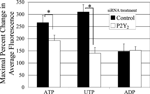
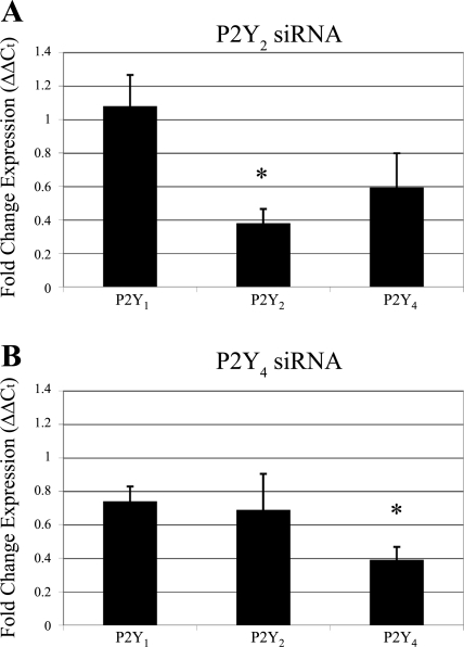
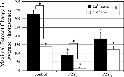
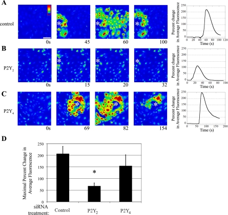
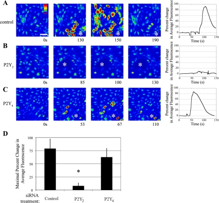
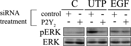
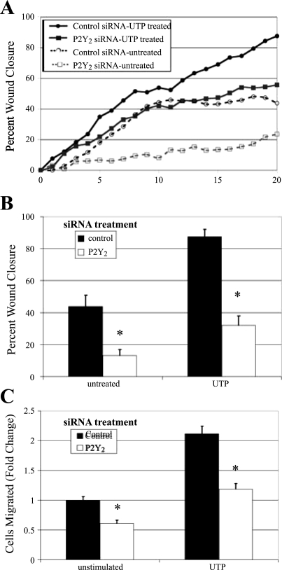
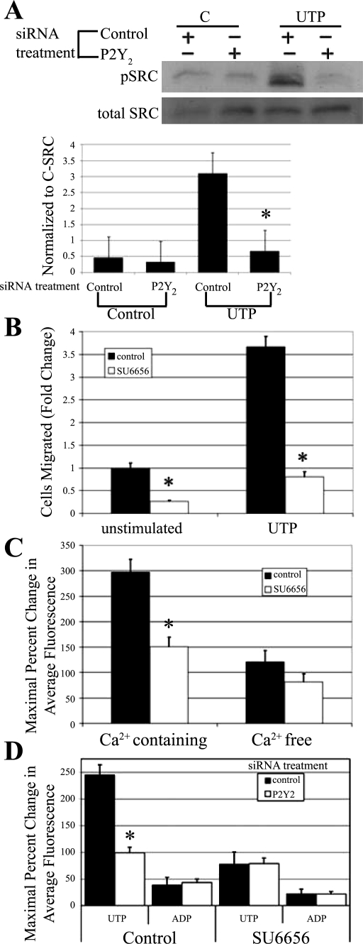
Injury to epithelial cells results in the release of ATP and stimulation of purinergic receptors and is thought to alter cell migration and wound repair. Medium from the injured cells triggers Ca(2+) mobilization and phosphorylation of ERK, both of which are inhibited if the medium is pretreated with apyrase. To understand the wound repair mechanism that occurs with injury, our goal was to determine which purinergic receptor(s) was the critical player in the wound response. We hypothesize that the P2Y(2) receptor is the key player in the response of corneal epithelial cells to cell damage and subsequent repair events. Cells transfected with short interfering RNA to either P2Y(2) or P2Y(4) were stimulated either by injury or addition of UTP and imaged using fluo 3-AM to monitor changes in fluorescence. When cells with downregulated P2Y(2) receptors were injured or stimulated with UTP, the intensity of the Ca(2+) release was reduced significantly. However, when cells with downregulated P2Y(4) receptors were stimulated, only the UTP-induced Ca(2+) response was reduced significantly. In addition, downregulation of the P2Y(2) receptor inhibited wound closure compared with unstimulated cells or cells transfected with nontargeting sequence. This downregulation resulted also in an attenuation in phosphorylation of Src and ERK. Together, these data indicate that the P2Y(2) receptor plays a major biological role in the corneal injury response and repair mechanisms.


Figures

Fig. 1.
Fig. 1.
P2Y2-receptor downregulation inhibits nucleotide induced Ca2+ response. HCLE cells were incubated in 5 μM fluo 3-AM for 30 min and imaged in a flow-through apparatus on a Zeiss LSM 510 confocal microscope. Cells were washed in HEPES-buffered saline for at least 30 s to establish baseline fluorescence. P2Y2-receptor short interfering RNA (siRNA)-transfected cells were stimulated with ATP (100 μM), UTP (100 μM), or ADP (250 μM) and compared with cells transfected with a nontargeting control siRNA. Maximal percent change in average fluorescence and SE of the mean were calculated. Graphs represent a minimum of six independent experiments. Significance was determined by Student's t-test: *P < 0.01.
Fig. 2.
Fig. 2.
Endogenous expression of P2Y receptors. Real-time PCR and the ΔΔCt (cycle threshold) method were used to determine expression of P2Y1, P2Y2, and P2Y4 receptors in epithelial cells. Data were normalized to the 18S ribosomal subunit and graphed as fold expression of confluency over subconfluency (30%) for each receptor. Each sample was run in triplicate on the same plate. Data are representative of a minimum of three independent experiments. Significance was determined by a Student's t-test.
Fig. 3.
Fig. 3.
Expression of P2Y receptors after transfection. Real-time PCR was performed to assay for expression of P2Y receptor mRNA after transfection. The ΔΔCt method was used to determine fold change expression of P2Y1, P2Y2, and P2Y4 receptors in cells transfected with P2Y2 (A) and P2Y4 (B) siRNA and compared with control cells transfected with a nontargeting siRNA sequence. Results were normalized to the 18S ribosomal subunit and graphed as relative expression compared with nontargeting control (normalized to 1). Data are representative of a minimum of six independent experiments. Significance compared with the nontargeting control was determined by a one-way ANOVA followed by Tukey's post hoc test: *P < 0.05.
Fig. 4.
Fig. 4.
P2Y2 and P2Y4 receptor downregulation decreases UTP-induced Ca2+ release. HCLE cells cultured on glass coverslips were incubated in 5 μM fluo 3-AM for 30 min and imaged in a flow-through apparatus on a Zeiss LSM 510 confocal microscope. Cells were washed in Ca2+-containing (solid bars) or Ca2+-free (open bars) HEPES-buffered saline for at least 30 s. P2Y2 and/or P2Y4 receptor siRNA transfected cells were stimulated with UTP (100 μM) and compared with cells transfected with a nontargeting control siRNA. Maximal percent change in average fluorescence and SE were calculated. Data are representative of at least three independent experiments. Significance of siRNA treated vs. control under the same Ca2+ condition was determined by a one-way ANOVA followed by Tukey's post hoc test: *P < 0.01. Significance of Ca2+ containing vs. Ca2+ free was determined by Student's t-test: P < 0.001.
Fig. 5.
Fig. 5.
P2Y2 receptor mediates in the injury-induced Ca2+ response in Ca2+-containing buffer. HCLE cells were incubated in 5 μM fluo 3-AM for 30 min and imaged in a flow-through apparatus on a Zeiss LSM 510 confocal microscope. Cells were washed in HEPES-buffered saline with Ca2+ for at least 30 s and stimulated by wounding. Intensity scale is shown in A, with red indicating highest Ca2+ levels and blue indicating lowest Ca2+ levels. The horizontal white bar in A represents 100 μm. AC: cells were transfected with the indicated siRNA sequence. Cells were washed in HEPES buffer containing Ca2+ and wounded. A series of images taken from a time course of a representative experiment of a wound (shown at asterisk) is presented, and percent change in average fluorescence was graphed (right). D: maximal percent change in average fluorescence was calculated and averaged for multiple repeats of the experiment. Data are representative of at least nine independent experiments. Significance was determined by a one-way ANOVA: *P < 0.01.
Fig. 6.
Fig. 6.
P2Y2 receptor mediates the injury-induced Ca2+ response in Ca2+-free buffer. HCLE cells were incubated in 5 μM fluo 3-AM for 30 min and imaged in a flow-through apparatus on a Zeiss LSM 510 confocal microscope. Cells were washed in Ca2+-free HEPES-buffered saline for at least 30 s and stimulated by wounding. Intensity scale is shown in A, with red indicating highest Ca2+ levels and blue indicating lowest Ca2+ levels. The horizontal white bar in A represents 100 μm. AC: cells were transfected with the indicated siRNA sequence. Cells were washed in Ca2+-free HEPES buffer and wounded. A series of images taken from a time course of a representative experiment of a wound (shown at asterisk) is presented, and percent change in average fluorescence was graphed (right). D: maximal percent change in average fluorescence was calculated and averaged for multiple repeats of the experiment. Data are representative of at least three independent experiments. Significance was determined by Student's t-test: *P < 0.05.
Fig. 7.
Fig. 7.
Purinergic receptor activation induces ERK phosphorylation. Cells were cultured on p60 culture dishes and transfected with an siRNA sequence against P2Y2 or nontargeting control siRNA (control). Cells in both groups were stimulated for 5 min with a media change (C), UTP (25 μM), or EGF (0.5 nM). Lysates of equivalent protein concentration were resolved using SDS-PAGE and immunoblotted with antibodies directed against total and phosphorylated ERK (pERK). Blots are representative of three independent experiments. Real-time PCR was performed to verify knockdown of P2Y2 receptor mRNA (data not shown). Data are representative of a minimum of six independent experiments.
Fig. 8.
Fig. 8.
P2Y2 receptor mediates cell migration. HCLE cells were transfected with siRNA against the P2Y2 receptor or a nontargeting control sequence. A: directed migration. Confluent cells in eight-well chamber slides were incubated overnight in media lacking growth factors. The media were replaced with either unsupplemented media or media containing 100 μM UTP immediately before wounding. Cells were placed on a heated microscope stage, wounded, and incubated in an environmental chamber at 37°C and 5% CO2 for 20 h. The wounds were demarcated, and contiguous regions were tiled and imaged every 20 min, and a representative run is graphed. B: percent wound closure from directed migration assays measured and averaged at 20 h. Data are representative of a minimum of three independent experiments. Significance was determined by a one-way ANOVA followed by Tukey's post hoc test: *P < 0.01. C: chemotactic migration. HCLEs were transfected with siRNA against the P2Y2 receptor or a nontargeting control sequence. Transwell migrations were performed for 8 h at 37°C with binding buffer alone (unstimulated) or binding buffer containing 1 μM UTP. Cells were stained with propidium iodide, counted in six randomly chosen fields (1.48 mm2), averaged, and normalized to unstimulated control, and SE of the mean was calculated. Data are representative of a minimum of three independent experiments. Significance was determined by Student's t-test: *P < 0.01.
Fig. 9.
Fig. 9.
Role of P2Y receptor regulation. A: HCLE cells were transfected with siRNA against the P2Y2 receptor or a nontargeting control sequence. Cells were incubated in the presence or absence of UTP for 5 min and immunoblotted with antibodies directed to pSrc and c-Src, and densitometric analysis was performed and normalized to c-Src. Control, nontargeting sequence; C, media change. Data are representative of a minimum of three independent experiments. Significance was determined by a Student's t-test: *P < 0.05. B: HCLE cells were incubated in the presence or absence of 10 μM Src kinase inhibitor SU6656 for the duration of the experiment. Transwell migrations were performed for 8 h at 37°C with binding buffer alone (unstimulated) or binding buffer containing 1 μM UTP. Cells were stained with propidium iodide, counted in six randomly chosen fields (1.46 mm2), averaged, and normalized to unstimulated control. Data are representative of a minimum of three independent experiments. Significance was determined by Student's t-test: *P < 0.01. C: HCLE cells were incubated in 5 μM fluo 3-AM in the presence or absence of 10 μM SU6656 for 30 min and imaged in a flow-through apparatus on a Zeiss LSM 510 confocal microscope. Cells were washed in either Ca2+-containing or Ca2+-free HEPES buffered saline ± 10 μM SU6656 for at least 30 s to obtain baseline. Cells were stimulated with UTP (100 μM), and maximal percent change in average fluorescence was calculated. Data are representative of a minimum of six independent experiments. Significance was determined by a Student's t-test: *P < 0.01. D: HCLE cells were transfected with siRNA against the P2Y2 receptor or a nontargeting control sequence. HCLE cells were incubated in 5 μM fluo 3-AM ± 10 μM SU6656 for 30 min and imaged in a flow-through apparatus on a Zeiss LSM 510 confocal microscope. Cells were washed in Ca2+-containing HEPES buffered saline ± 10 μM SU6656 for at least 30 s. Cells were stimulated with ADP (100 μM), washed, and stimulated with UTP (100 μM), and maximal percent change in response to each agonist in average fluorescence was calculated. Data are representative of a minimum of six independent experiments. Significance was determined by a Student's t-test: *P < 0.01.

References

    1. Abbracchio MP, Burnstock G. Purinergic signalling: pathophysiological roles. Jpn J Pharmacol 78: 113–145, 1998 - PubMed
    1. Abbracchio MP, Burnstock G, Boeynaems JM, Barnard EA, Boyer JL, Kennedy C, Knight GE, Fumagalli M, Gachet C, Jacobson KA, Weisman GA. International Union of Pharmacology LVIII: Update on the P2Y G protein-coupled nucleotide receptors: from molecular mechanisms and pathophysiology to therapy. Pharmacol Rev 58: 281–341, 2006. - PMC - PubMed
    1. Arthur DB, Georgi S, Akassoglou K, Insel PA. Inhibition of apoptosis by P2Y2 receptor activation: novel pathways for neuronal survival. J Neurosci 26: 3798–3804, 2006 - PMC - PubMed
    1. Arthur DB, Akassoglou K, Insel PA. P2Y2 and TrkA receptors interact with Src family kinase for neuronal differentiation. Biochem Biophys Res Commun 347: 678–682, 2006 - PubMed
    1. Berridge MJ. Inositol trisphosphate and diacyglycerol: two interacting second messengers. Annu Rev Biochem 56: 159–193, 1987 - PubMed

Publication types

Substances