. 2010 Apr 28;2(29):29ra31.
doi: 10.1126/scitranslmed.3000928.

Cystic fibrosis pigs develop lung disease and exhibit defective bacterial eradication at birth

Affiliations

Cystic fibrosis pigs develop lung disease and exhibit defective bacterial eradication at birth

David A Stoltz et al. Sci Transl Med. .

Abstract

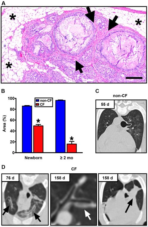
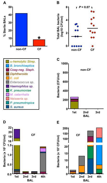
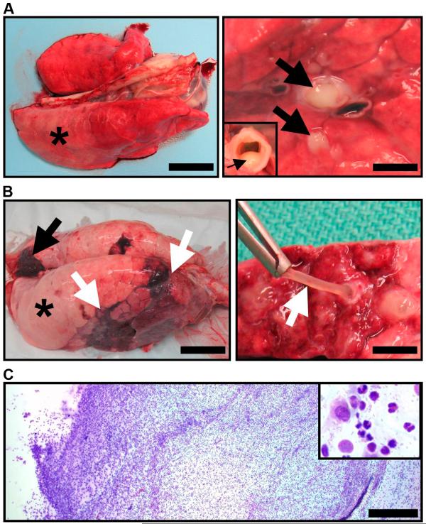
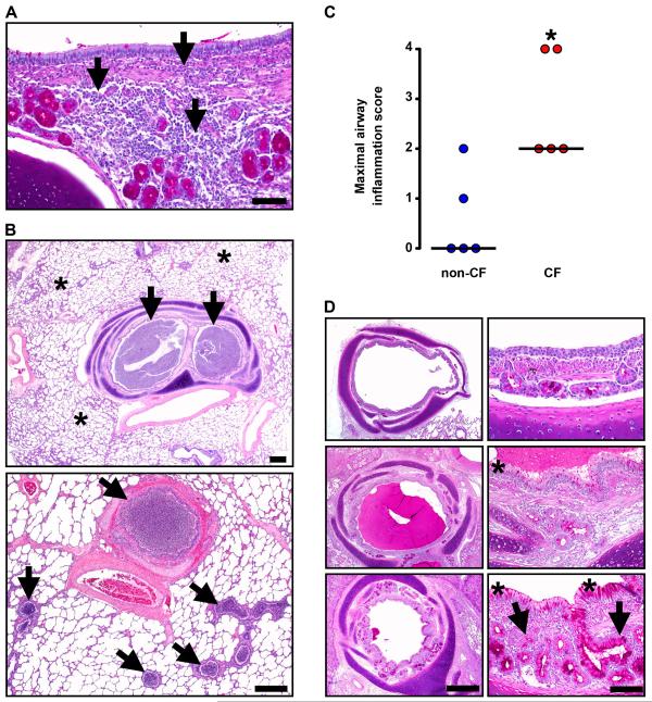
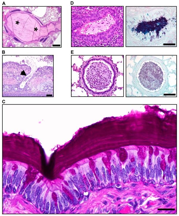
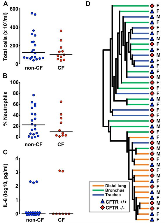
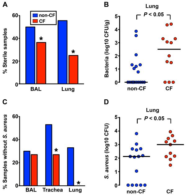
Lung disease causes most of the morbidity and mortality in cystic fibrosis (CF). Understanding the pathogenesis of this disease has been hindered, however, by the lack of an animal model with characteristic features of CF. To overcome this problem, we recently generated pigs with mutated CFTR genes. We now report that, within months of birth, CF pigs spontaneously developed hallmark features of CF lung disease, including airway inflammation, remodeling, mucus accumulation, and infection. Their lungs contained multiple bacterial species, suggesting that the lungs of CF pigs have a host defense defect against a wide spectrum of bacteria. In humans, the temporal and causal relations between inflammation and infection have remained uncertain. To investigate these processes, we studied newborn pigs. Their lungs showed no inflammation but were less often sterile than controls. Moreover, after introduction of bacteria into their lungs, pigs with CF failed to eradicate bacteria as effectively as wild-type pigs. These results suggest that impaired bacterial elimination is the pathogenic event that initiates a cascade of inflammation and pathology in CF lungs. Our finding that pigs with CF have a host defense defect against bacteria within hours of birth provides an opportunity to further investigate CF pathogenesis and to test therapeutic and preventive strategies that could be deployed before secondary consequences develop.


Figures

Fig. 1
Fig. 1. CF pigs develop pancreatic and pulmonary disease
(A) Pancreas from Case #1. Adipose tissue (asterisks) surrounded degenerative exocrine lobules, large obstructed ducts and fibrosis (arrows), HE stain, scale bar, 240 μm. (B) Lobular exocrine tissue in newborn pigs and pigs greater than 2 months age (data from newborn pigs was previously published in (25)). Low magnification images of pancreatic parenchyma and interlobular connective tissue were quantified, and lobular tissue area was recorded as a percent of the total area for each image (*, P < 0.01, Mann-Whitney test). Chest X-ray computed tomography from (C) non-CF and (D) CF pigs (Case #5, left; Case #4, middle and right). Airway wall thickening (arrows) appeared with time in CF pigs. At 158 d, obstruction of tracheal bronchus was observed in Case #4 with collapse of the associated lung segment (black arrow).
Fig. 2
Fig. 2. CF pig lungs contain bacteria
Data are from BAL, with several animals having multiple lavages at 4-6 week intervals. (A) Percentage of sterile BAL liquid samples obtained over time from non-CF and CF animals. * P < 0.05. (B) Number of bacteria recovered in tracheal lobe BAL samples obtained over time from non-CF and CF animals. Horizontal line denotes median. n=7 non-CF and n=5 CF animals. Each data point represents a BAL sample from an animal. Several animals had sequential BAL over time and are represented more than once. (C-E) Serial BAL cultures from a non-CF pig (C) and two CF pigs (Case #5, D; Case #4, E). Data are color-coded to indicate individual species of bacteria.
Fig. 3
Fig. 3. CF pigs spontaneously develop lung disease
(A) Lung, Case #1. Right caudal lung did not collapse (*)(scale bar, 3.4 cm) and sectioning revealed purulent material in obstructed airways (arrows)(scale bar, 2.9 mm) and tracheal lumen (arrow, inset). (B) Lung, Case #4. Lungs had atelectasis (black arrow), hyperinflation (asterisk) and pneumonia (white arrows, left panel, scale bar, 5 cm). Airways were obstructed by tenacious and resilient mucus (white arrow, right panel, scale bar, 6.6 mm). (C) Mucus cytology, Case #4. Mucus had a high cellularity composed primarily of degenerative neutrophils (inset) with macrophages and bacteria (scale bar, 0.4 mm).
Fig. 4
Fig. 4. Lung disease in CF pigs replicates that in human CF
(A) Case #3. Infiltration of the airway wall by lymphocytes and plasma cells (arrows), PAS stain, scale bar, 80 μm. (B) Case #1. Obstruction (arrows) of bronchi and small bronchioles was a striking feature in otherwise unaffected lung (asterisks). HE stain, scale bars, 0.75 mm. (C) Airways from non-CF and CF pigs were scored for the most severe degree of leukocytic infiltration detected with the following scale: 0 – no leukocytes detected; 1 – rare individual to scattered leukocytes occasionally seen in airway wall with lack of cellular aggregates; 2 – grade 1 plus minor to moderate leukocyte aggregates detected in wall ± rare in lumen; 3 – grade 2 plus in multiple airways, with early wall injury, and luminal leukocytic aggregates; and 4 – grade 3, plus airway wall destruction, leukocytes filling and obstructing airways. For grades 1 and 2, leukocytes were primarily lymphocytes and plasma cells, and for grades 3 and 4, leukocytes were primarily neutrophils. Each point represents a sample from a different animal. CF pigs airways had more severe scores of airway infiltration (*P <0.05, Mann-Whitney, horizontal line indicates the median). (D) Case #4. Airways (left panels, scale bar, 0.7 mm) ranged from relatively unaffected (top) to severe disease (bottom, note that luminal mucocellular plug was removed at necropsy) with airway wall thickening. Surface epithelium (right panels, scale bar, 70 μm) ranged from near normal (top) to mucinous and hyperplastic change (asterisks) in moderate to severe disease (middle and bottom panels). Note that hypertrophy/hyperplasia of submucosal glands (arrows, bottom right) was uncommon and generally restricted to the most severe and chronically affected airways.
Fig. 5
Fig. 5. Mucus accumulates in CF airways
(A) Case #4. Some airways were completely obstructed by mucocellular material (asterisks, HE stain, scale bar, 0.44 mm). (B) Case #1. Submucosal gland ducts were rarely dilated with stringy mucocellular debris (arrow, HE stain, scale bar, 75 μm). (C) Case #1. Some bronchial walls were lined by mucus with a distinct lamellar appearance (PAS stain, scale bar, 22 μm). (D) Bronchioles, Case #1. An obstructed bronchiole (which lack submucosal glands) with proliferative and mucinous changes in surface epithelium, HE (left panel) and ABPY (right) stains, scale bar, 71 μm. (E) Bronchioles, Case #1. Some obstructed bronchioles lacked proliferative and mucinous changes to epithelium, HE (left) and ABPY (right) stains, scale bar, 71 μm.
Fig. 6
Fig. 6. At birth, CF pig lungs lack inflammation
(A) Total cell counts, (B) Neutrophil percentages, and (C) IL-8 levels in BAL liquid obtained from non-CF and CF piglets within 6-12 h following birth. Each point represents a sample from a different animal. Horizontal line denotes median. No statistically significant differences were observed between non-CF and CF BAL liquid total cell counts, neutrophil percentages, or IL-8 levels. (D) Unsupervised hierarchical clustering of mRNA samples from trachea, bronchus, and distal lung samples obtained from newborn piglets (6-12 h old). Samples segregated into two clusters: airways (trachea and bronchus) and distal lung, but there was no clustering based on CFTR genotype. M and F refer to male and female pigs.
Fig. 7
Fig. 7. Newborn CF piglets fail to eradicate bacteria
(A) Percentage of BAL liquid and lung homogenate samples that were sterile (no culturable bacteria) from newborn non-CF and CF pigs. BAL liquid n=20 non-CF and n=11 CF; lung homogenate n=18 non-CF and n=12 CF. Data are from 6 litters. * P < 0.05. (B) Quantitative microbiology on lung homogenate tissue samples. 6-12 h-old piglets were euthanized and lungs were sterilely removed. Each point represents an individual animal. Horizontal line denotes median. (C) Percentage of BAL liquid, trachea, and lung homogenate samples that contained no S. aureus. Samples were obtained from newborn non-CF and CF piglets after intrapulmonary challenge with S. aureus. (* P < 0.05 Chi-square test). (D) Bacteria recovered from tracheal lobe after S. aureus intrapulmonary challenge. Each point represents a different animal. Horizontal line denotes median. Data are from 5 litters. n=15 non-CF and n=11 CF.

References

    1. Riordan JR, et al. Identification of the cystic fibrosis gene: cloning and characterization of complementary DNA. Science. 1989;245:1066–1073. - PubMed
    1. Welsh MJ, Ramsey BW, Accurso F, Cutting GR. Cystic Fibrosis. In: Scriver CR, et al., editors. The Metabolic and Molecular Basis of Inherited Disease. McGraw-Hill; New York: 2001. pp. 5121–5189.
    1. Quinton P. Physiological basis of cystic fibrosis: a historical perspective. Physiol. Rev. 1999;79(Suppl. 1):S3–S22. - PubMed
    1. Rowe SM, Miller S, Sorscher EJ. Cystic fibrosis. N. Engl. J. Med. 2005;352(19):1992–2001. - PubMed
    1. Guggino WB. Cystic fibrosis and the salt controversy. Cell. 1999;96(5):607–610. - PubMed

Publication types

MeSH terms