. 2010 Aug;78(3):310-7.
doi: 10.1038/ki.2010.131. Epub 2010 Apr 28.

Renal histopathology and crystal deposits in patients with small bowel resection and calcium oxalate stone disease

Affiliations

Renal histopathology and crystal deposits in patients with small bowel resection and calcium oxalate stone disease

Andrew P Evan et al. Kidney Int. 2010 Aug.

Abstract

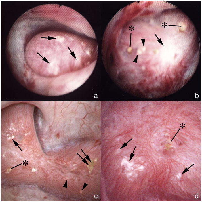
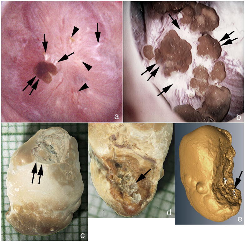
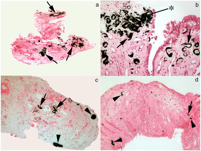
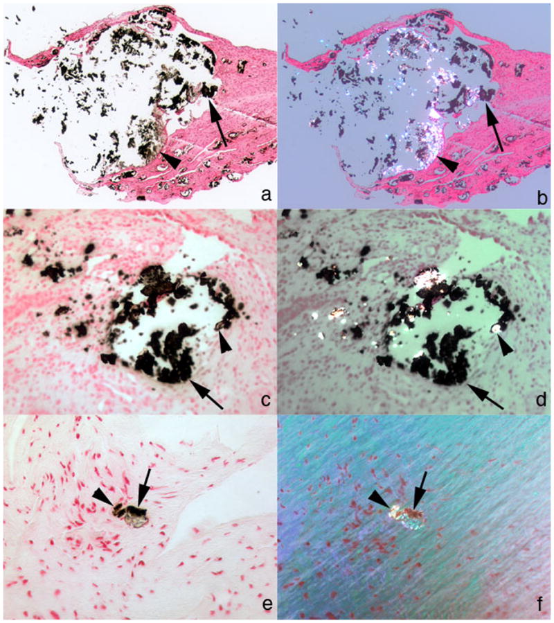
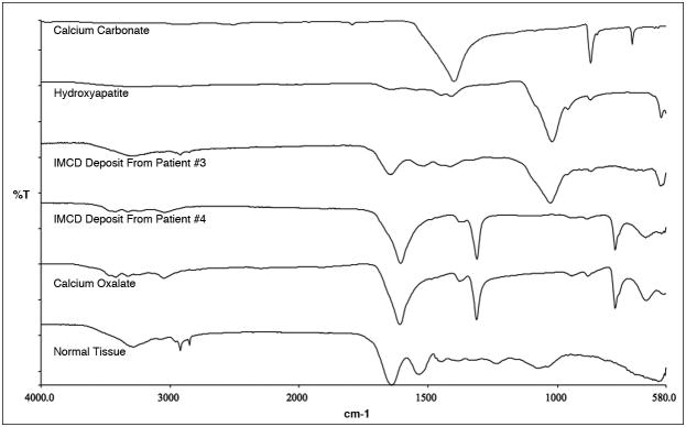
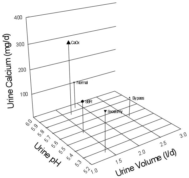
We present here the anatomy and histopathology of kidneys from 11 patients with renal stones following small bowel resection, including 10 with Crohn's disease and 1 resection in infancy for unknown cause. They presented predominantly with calcium oxalate stones. Risks of formation included hyperoxaluria (urine oxalate excretion greater than 45 mg per day) in half of the cases, and acidic urine of reduced volume. As was found with ileostomy and obesity bypass, inner medullary collecting ducts (IMCDs) contained crystal deposits associated with cell injury, interstitial inflammation, and papillary deformity. Cortical changes included modest glomerular sclerosis, tubular atrophy, and interstitial fibrosis. Randall's plaque (interstitial papillary apatite) was abundant, with calcium oxalate stone overgrowth similar to that seen in ileostomy, idiopathic calcium oxalate stone formers, and primary hyperparathyroidism. Abundant plaque was compatible with the low urine volume and pH. The IMCD deposits all contained apatite, with calcium oxalate present in three cases, similar to findings in patients with obesity bypass but not an ileostomy. The mechanisms for calcium oxalate stone formation in IMCDs include elevated urine and presumably tubule fluid calcium oxalate supersaturation, but a low calcium to oxalate ratio. However, the mechanisms for the presence of IMCD apatite remain unknown.


Conflict of interest statement

Disclosure:

All the authors declared no competing interests.

Figures

Figure 1
Figure 1. Coexistence of attached stones and BD plugging on the same papillae
(a) Large area of white interstitial (Randall’s) plaque (single arrows) on one papilla. (b) In another patient a large area of white plaque (single arrow) is seen in intermixed with areas of yellow plaque (IMCD crystal deposits, arrowheads) and crystalline plugs (asterisks) protruding from dilated BD. (c) A papilla from another patient shows a smaller area of white plaque (single arrow) intermixed with yellow plaque (arrowheads), BD plugs (asterisk) and an attached stone (double arrow). (d) At higher magnification, a dilated BD is seen with a protruding crystal plug (asterisk) near several small areas of white plaque (single arrows).
Figure 2
Figure 2. Details of attached stones
(a) An attached stone (double arrow) is seen resting on a region of white plaque (single arrows) and intermixed with small areas of white (single arrow) and yellow plaque (arrowheads). Patient 8 had numerous attached stones (double arrow) atop an extensive area of white plaque (single arrows) much like that found in some ICSF. Analysis of attached stones by μ-CT revealed these to be composed of primarily of CaOx with small sites of apatite corresponding to a site of attachment to white (Randall’s) plaque. (c & d) g Light microscopic image of an attached revealing the smooth urinary (c) and papillary (d) surface morphology. The papillary surface (d) shows a concave region with crystalline material (single arrow) consistent with an attachment site. The urinary surface (c) shows a damaged region (double arrow) generated during stone removal. (e) Reconstruction of μ-CT images shows regions of CaOx in yellow and areas of apatite in white. The white regions are appear to present the attachment site.
Figure 3
Figure 3. Relative densities of interstitial plaque and IMCD deposits
(a,b) Large areas of interstitial plaque (Randall’s) (arrows) are seen surrounding the thin loops of Henle and extending to the base of the urothelial cells (asterisk). (c,d) Areas of interstitial plaque (arrows) and plugged IMCD (arrowheads) are found in the same biopsy sample at varying amounts. None of these IMCD deposits contained birefringent crystals. Interstitial fibrosis was associated with IMCD plugs. Original magnification × 100 (a,b); × 50 (c,d).
Figure 4
Figure 4. IMCD deposits mixture of apatite and CaOx
(a–d) Two different large IMCD plugs from separate patients are seen under non-polarizing (a, c) vs polarizing (b, d) optics. These deposits show birefringent (arrowheads) and non-birefringent (single arrows) crystals forming the same deposit yet not admixed. The non-birefringent crystals are probably apatite and the birefringent crystals CaOx. (e, f) An occasional small IMCD plug also possessed both birefringent (see arrowheads in panels e, f) and non-birefringent (see single arrows in panels e, f)) crystals. Original magnification × 25 (a–d); × 100 (e,f).
Figure 5
Figure 5. Micro-Fourier-Transform Infrared spectrometer analysis of IMCD deposits
IMCD deposits show a spectral band matching that of the hydroxyapatite and calcite standards in patient #3 and CaOx standard in patient #4. Tissue with embedding medium, a control, displays bands in common with the three standards.
Figure 6
Figure 6. Principle determinants of interstitial plaque
Normal subjects (Normal) and idiopathic CaOx stone formers (CaOx) have urine pH (y-axis) values near 6 as well as similar urine volumes (x-axis), but the stone formers have much higher calcium excretion (z-axis height) and abundant plaque (size of symbol) vs. small amounts for normals. Bypass patients (Bypass) have normal amounts of plaque; low urine calcium and high volume offset the low urine pH. Small bowel resection (SBR) and ileostomy patients (Ileostomy) have low urine pH and volume, and normal calcium excretions, and both form abundant plaque.

References

    1. Evan AP, Lingeman JE, Coe FL, et al. Randall’s plaque of patients with nephrolithiasis begins in basement membranes of thin loops of Henle. J Clin Invest. 2003;111:607–616. - PMC - PubMed
    1. Evan AP, Coe FL, Gillen D, et al. Renal intratubular crystals and hyaluronan staining occur in stone formers with bypass surgery but not with idiopathic calcium oxalate stones. Anat Rec (Hoboken) 2008;291:325–334. - PubMed
    1. Evan AP, Lingeman JE, Coe FL, et al. Intra-tubular deposits, urine and stone composition are divergent in patients with ileostomy. Kidney Int. 2009;76:1081–1088. - PMC - PubMed
    1. Miller NL, Gillen DL, Williams JC, Jr, et al. A formal test of the hypothesis that idiopathic calcium oxalate stones grow on Randall’s plaque. BJU Int. 2009;103:966–971. - PMC - PubMed
    1. Evan AE, Lingeman JE, Coe FL, et al. Histopathology and surgical anatomy of patients with primary hyperparathyroidism and calcium phosphate stones. Kidney Int. 2008;74:223–229. - PubMed

Publication types

MeSH terms