

Cell-based osteoprotegerin therapy for debris-induced aseptic prosthetic loosening on a murine model
- PMID: 20428210
- PMCID: PMC2914841
- DOI: 10.1038/gt.2010.64
Cell-based osteoprotegerin therapy for debris-induced aseptic prosthetic loosening on a murine model
Abstract
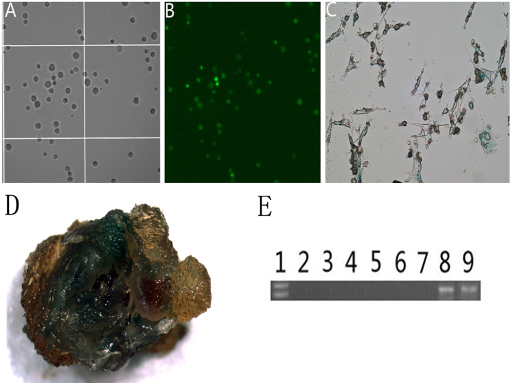
Exogenous osteoprotegerin (OPG) gene modification appears a therapeutic strategy for osteolytic aseptic loosening. The feasibility and efficacy of a cell-based OPG gene delivery approach were investigated using a murine model of knee prosthesis failure. A titanium pin was implanted into mouse proximal tibia to mimic a weight-bearing knee arthroplasty, followed by titanium particles challenge to induce periprosthetic osteolysis. Mouse fibroblast-like synoviocytes were transduced in vitro with either AAV-OPG or AAV-LacZ before transfused into the osteolytic prosthetic joint 3 weeks post surgery. Successful transgene expression at the local site was confirmed 4 weeks later after killing. Biomechanical pullout test indicated a significant restoration of implant stability after the cell-based OPG gene therapy. Histology revealed that inflammatory pseudo-membranes existed ubiquitously at bone-implant interface in control groups, whereas only observed sporadically in OPG gene-modified groups. Tartrate-resistant acid phosphatase+osteoclasts and tumor necrosis factor α, interleukin-1β, CD68+ expressing cells were significantly reduced in periprosthetic tissues of OPG gene-modified mice. No transgene dissemination or tumorigenesis was detected in remote organs and tissues. Data suggest that cell-based ex vivo OPG gene therapy was comparable in efficacy with in vivo local gene transfer technique to deliver functional therapeutic OPG activities, effectively halted the debris-induced osteolysis and regained the implant stability in this model.
Conflict of interest statement
The authors declare no conflict of interest.
Figures


References
-
- Holt G, Murnaghan C, Reilly J, Meek RM. The biology of aseptic osteolysis. Clin Orthop Relat Res. 2007;460:240–252. - PubMed
-
- Purdue PE, Koulouvaris P, Potter HG, Nestor BJ, Sculco TP. The cellular and molecular biology of periprosthetic osteolysis. Clin Orthop Relat Res. 2007;454:251–261. - PubMed
-
- Suzuki Y, Nishiyama T, Hasuda K, Fujishiro T, Niikura T, Hayashi S, et al. Effect of etidronate on COX-2 expression and PGE(2) production in macrophage-like RAW 264.7 cells stimulated by titanium particles. J Orthop Sci. 2007;12:568–577. - PubMed
-
- Kim KJ, Chiba J, Rubash HE. In vivo and in vitro analysis of membranes from hip prostheses inserted without cement. J Bone Joint Surg Am. 1994;76:172–180. - PubMed
-
- Shanbhag AS, Jacobs JJ, Black J, Galante JO, Glant TT. Cellular mediators secreted by interfacial membranes obtained at revision total hip arthroplasty. J Arthroplasty. 1995;10:498–506. - PubMed
Publication types
MeSH terms
Substances
Grants and funding
LinkOut - more resources
Full Text Sources
