. 2010 Sep;17(9):1162-74.
doi: 10.1038/gt.2010.56. Epub 2010 Apr 29.

rAAV2/5 gene-targeting to rods:dose-dependent efficiency and complications associated with different promoters

Affiliations

rAAV2/5 gene-targeting to rods:dose-dependent efficiency and complications associated with different promoters

W A Beltran et al. Gene Ther. 2010 Sep.

Abstract

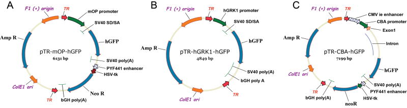
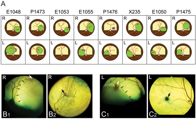
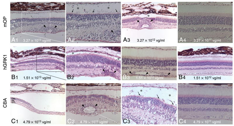
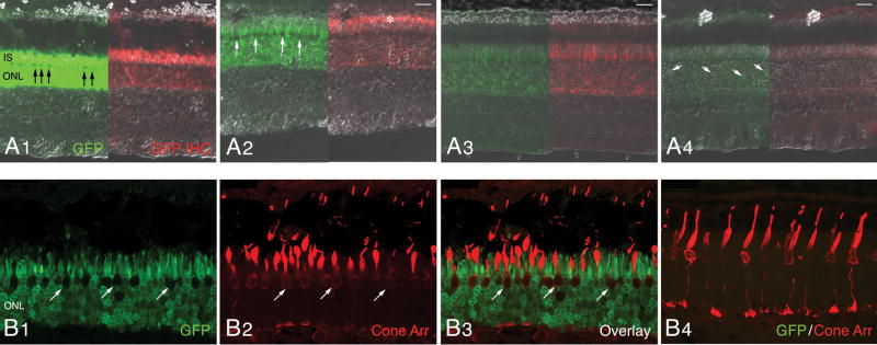
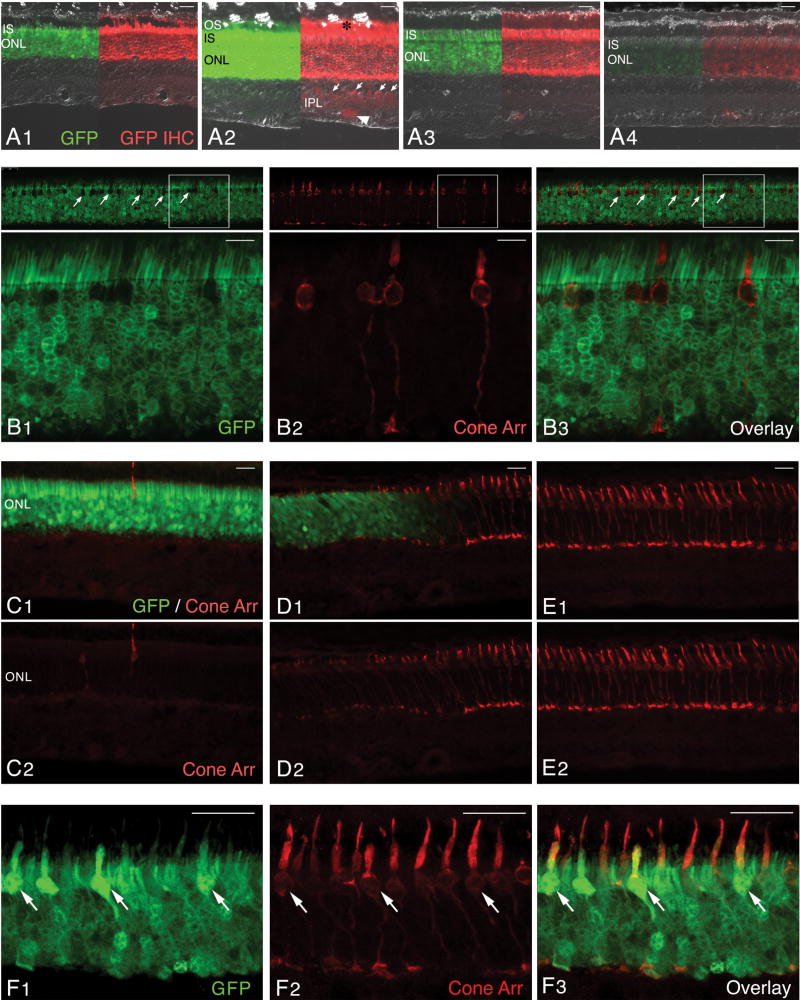
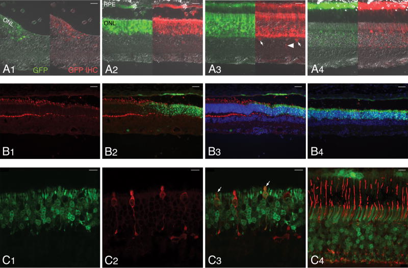
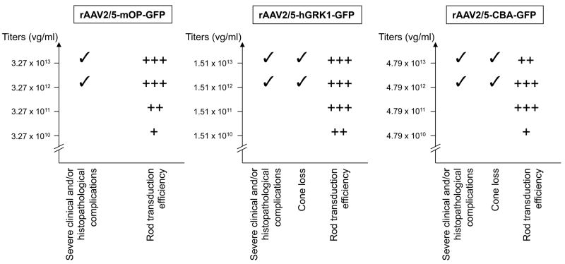
A prerequisite for using corrective gene therapy to treat humans with inherited retinal degenerative diseases that primarily affect rods is to develop viral vectors that target specifically this population of photoreceptors. The delivery of a viral vector with photoreceptor tropism coupled with a rod-specific promoter is likely to be the safest and most efficient approach to target expression of the therapeutic gene to rods. Three promoters that included a fragment of the proximal mouse opsin promoter (mOP), the human G-protein-coupled receptor protein kinase 1 promoter (hGRK1), or the cytomegalovirus immediate early enhancer combined with the chicken β actin proximal promoter CBA were evaluated for their specificity and robustness in driving GFP reporter gene expression in rods, when packaged in a recombinant adeno-associated viral vector of serotype 2/5 (AAV2/5), and delivered via subretinal injection to the normal canine retina. Photoreceptor-specific promoters (mOP, hGRK1) targeted robust GFP expression to rods, whereas the ubiquitously expressed CBA promoter led to transgene expression in the retinal pigment epithelium, rods, cones and rare Müller, horizontal and ganglion cells. Late onset inflammation was frequently observed both clinically and histologically with all three constructs when the highest viral titers were injected. Cone loss in the injected regions of the retinas that received the highest titers occurred with both the hGRK1 and CBA promoters. Efficient and specific rod transduction, together with preservation of retinal structure was achieved with both mOP and hGRK1 promoters when viral titers in the order of 10(11)vg ml(-1) were used.


Conflict of interest statement

CONFLICT of INTEREST: WWH and the University of Florida have a financial interest in the use of rAAV therapies, and own equity in a company (AGTC Inc.) that might, in the future, commercialize some aspects of this work. All remaining authors declare that they have no competing financial interests.

Figures

Figure 1
Figure 1. Schematic diagrams of the plasmid constructs used to produce rAAV
(A) Map of the pTR-mOP-hGFP plasmid DNA used to make the rAAV2/5-mOP-GFP virus. (B) Map of the pTR-hGRK1-hGFP plasmid DNA used to make the rAAV2/5-hGRK1-GFP virus. (C) Map of the pTR-CBA-hGFP plasmid DNA used to make the rAAV2/5-CBA-GFP virus. TR represents AAV2 inverted terminal repeats; mOP represents a fragment of the proximal mouse opsin promoter; hGRK1 represents a fragment of the human G-protein-coupled receptor protein kinase 1 promoter; CMV ie represents the cytomegalovirus immediate early enhancer; CBA represents the chicken beta-actin promoter; SV40 SD/SA represents the SV40 late viral protein gene 16S/19S splice donor and acceptor signal; hGFP represents the coding sequence of the humanized green fluorescence protein gene; SV40 (poly A) and bGH poly (A) represent polyadenylation signals; HSV-tk represents the thymidine kinase promoter of the herpes simplex virus, and Neo R represents the coding sequence of the neomycin resistance gene.
Figure 2
Figure 2. Location and extent of subretinal injections
(A) Diagrams of the fundus of the 16 eyes in which a subretinal injection was attempted. The green areas show the locations where subretinal blebs were achieved. The dark spot within the area of the subretinal bleb shows the location of the retinotomy. Purple areas show the location where some viral solution was seen injected under the RPE. [Note: the canine fundus is comprised of a tapetal region (superior; labeled in yellow; RPE non pigmented) and a non-tapetal region (inferior; labeled in brown; RPE pigmented)]. (B1–2) Fundus photographs of the right eye of dog X235 immediately after performing the subretinal injection (B1) and at termination (day 56; B2). (C1–2) Fundus photographs of the left eye of dog X235 immediately after performing the subretinal injection (C1) and at termination (day 56; C2). Asterisks show the same retinal location at time of injection and termination; small black arrows point to the edge of the subretinal bleb; large white arrows show the retinotomy site (at time of injection), and large black arrows point to the subsequent scar visible at termination.
Figure 3
Figure 3. Histopathological findings following subretinal injection with three different rAAV2/5 constructs
(A1–4) H&E stained retinal section of eyes treated with different titers of rAAV2/5-mOP-GFP: E1048-R (3.27 × 10¹³ vg/ml) (A1–2); E1048-L (3.27 × 10¹² vg/ml) (A3); and P1473-R (3.27 × 10¹¹ vg/ml)) (A4). (B1–4) H&E stained retinal section of eyes treated with different titers of rAAV2/5-hGRK1-GFP: E1055-R (1.51 × 10¹³ vg/ml) (B1–2); P1476-L (1.51 × 10¹² vg/ml) (B3); and E1055-L (1.51 × 10¹² vg/ml) (B4). (C1–4) H&E stained retinal section of eyes treated with different titers of rAAV2/5-CBA-GFP: E1050-R (4.79 × 10¹³ vg/ml) (C1); E1050-L (4.79 × 10¹² vg/ml) (C2–3); P1475-R (4.79 × 10¹¹ vg/ml) (C4). Abbreviations: ONL, outer nuclear layer; INL, inner nuclear layer; GCL, ganglion cell layer. Scale bars: (A1, A3, A4, B1, B4, C2) 40 μm; (B3, C4) 20 μm; (C1) 80 μm.
Figure 4
Figure 4. Transduction pattern of AAV2/5-mOP-GFP following subretinal delivery to the normal canine retina
Native GFP fluorescence (green) and GFP immunolabeling (red) images merged with DIC/Nomarski optics; images show the GFP expression with decreasing viral vector titers (A1–4). A1) Retina of E1048-R (3.27 × 10¹³ vg/ml). A2) Retina of E1048-L (3.27 × 10¹² vg/ml). A3) Retina of P1473-R (3.27 × 10¹¹ vg/ml). A4) Retina of P1473-L (3.27 × 10¹⁰ vg/ml). Confocal microscopy images of native GFP (green) and cone arrestin (red) immunolabeling in the injected area (B1–3) and non-injected area (B4) of retina E1048-R. Abbreviations: IS, inner segments; ONL, outer nuclear layer; GFP, green fluorescent protein; IHC, immunohistochemistry; Cone Arr. cone arrestin. Scale bars: (A14) 20 μm; (B14) 25 μm.
Figure 5
Figure 5. Transduction pattern of AAV2/5-hGRK1-GFP following subretinal delivery to the normal canine retina
(A1–4) Native GFP fluorescence (green) and GFP immunolabeling (red) images merged with DIC/Nomarski optics; images show the GFP expression with decreasing viral vector titers. (A1) Retina of E1055-R (1.51 × 10¹³ vg/ml). (A2) Retina of P1476-L (1.51 × 10¹² vg/ml). (A3) Retina of X235-R (1.51 × 10¹¹ vg/ml). (A4) Retina of X235-L (1.51 × 10¹⁰ vg/ml). (B1–3) Confocal microscopy images of native GFP (green) and cone arrestin (red) immunolabeling in the injected area of retina P1476-L.(C–E) Epifluorescence microscopy images of native GFP (green) and cone arrestin (red) immunolabeling in the center (C1–2), at the border (D1–2), and outside the injected retinal area of E1055-R. (F1–3) Confocal microscopy images of native GFP (green) and cone arrestin (red) immunolabeling in the injected area of retina E1055-R. Abbreviations: OS, outer segments; IS, inner segments; ONL, outer nuclear layer; INL; GFP, green fluorescent protein; IHC, immunohistochemistry; Cone Arr. cone arrestin. Scale bars: (A14; C12, D12, E12) 20 μm; (B13) 10 μm; (F13) 25 μm.
Figure 6
Figure 6. Transduction pattern of AAV2/5-CBA-GFP following subretinal delivery to the normal canine retina
Native GFP fluorescence (green) and GFP immunolabeling (red) images merged with DIC/Nomarski optics; images show the GFP expression with decreasing viral vector titers (A1–4). A1) Retina of E1050-R (4.79 × 10¹³ vg/ml). A2) Retina of E1050-L (4.79 × 10¹² vg/ml). A3) Retina of P1475-R (4.79 × 10¹¹ vg/ml). A4) Retina of P1475-L (4.79 × 10¹⁰ vg/ml). Epifluorescence microscopy images at the border (B1–3) and in the center (B4) of the injected retinal area of E1050-L (4.79 × 10¹² vg/ml). Confocal microscopy images of native GFP (green) and cone arrestin (red) immunolabeling in the injected area of retina E1050-L (4.79 × 10¹² vg/ml)(C1–3), and retina P1475-R (4.79 × 10¹¹ vg/ml) (C4). Abbreviations: RPE, retinal pigment epithelium, ONL, outer nuclear layer; GFP, green fluorescent protein; IHC, immunohistochemistry; Cone Arr. cone arrestin. Scale bars: (A14) 20 μm; (B14) 40 μm; (C14) 10 μm.
Figure 7
Figure 7
Summary of rod transduction efficiency and complications observed with the three viral vector constructs evaluated in this study. (A) rAAV2/5-mOP-GFP; (B) rAAV2/5-hGRK1-GFP; (C) rAAV2/5-CBA-GFP. +: limited; ++: moderate; +++: intense.

References

    1. Acland GM, Aguirre GD, Ray J, Zhang Q, Aleman TS, Cideciyan AV, et al. Gene therapy restores vision in a canine model of childhood blindness. Nat Genet. 2001;28(1):92–95. - PubMed
    1. Pang JJ, Chang B, Kumar A, Nusinowitz S, Noorwez SM, Li J, et al. Gene therapy restores vision-dependent behavior as well as retinal structure and function in a mouse model of RPE65 Leber congenital amaurosis. Mol Ther. 2006;13(3):565–572. - PubMed
    1. Weber M, Rabinowitz J, Provost N, Conrath H, Folliot S, Briot D, et al. Recombinant adeno-associated virus serotype 4 mediates unique and exclusive long-term transduction of retinal pigmented epithelium in rat, dog, and nonhuman primate after subretinal delivery. Mol Ther. 2003;7(6):774–781. - PubMed
    1. Bainbridge JW, Smith AJ, Barker SS, Robbie S, Henderson R, Balaggan K, et al. Effect of gene therapy on visual function in Leber’s congenital amaurosis. N Engl J Med. 2008;358(21):2231–2239. - PubMed
    1. Maguire AM, Simonelli F, Pierce EA, Pugh EN, Jr, Mingozzi F, Bennicelli J, et al. Safety and efficacy of gene transfer for Leber’s congenital amaurosis. N Engl J Med. 2008;358(21):2240–2248. - PMC - PubMed

Publication types