. 2010 Apr 23;5(4):e10336.
doi: 10.1371/journal.pone.0010336.

Temporal characterization of homology-independent centromere coupling in meiotic prophase

Affiliations

Temporal characterization of homology-independent centromere coupling in meiotic prophase

David Obeso et al. PLoS One. .

Abstract

Background: Over the past thirty years several reports of the pairing or association of non-homologous centromeres during meiotic prophase have appeared in the literature. Recently, the homology-independent pairwise association of centromeres, termed centromere coupling, was also reported in budding yeast. It seems paradoxical that centromeres would pair with non-homologous partners during a process intended to align homologous chromosomes, yet the conservation of this phenomenon across a wide range of species suggests it may play an important role in meiosis.

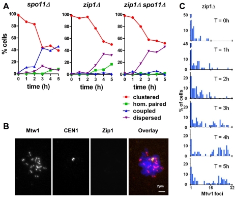
Principal findings: To better define the role of this phenomenon in budding yeast, experiments were preformed to place centromere coupling within the context of landmark meiotic events. Soon after the initiation of the meiotic program, centromeres were found to re-organize from a single cluster into non-homologous couples. Centromere coupling is detected as soon as chromosome replication is finished and persists while the recombination protein Dmc1 is loaded onto the chromosomes, suggesting that centromere coupling persists through the time of double strand break formation. In the absence of the synaptonemal complex component, Zip1, centromere coupling was undetectable, at all times examined, confirming the essential role of this protein on this process. Finally, the timely release of centromere coupling depends on the recombination-initiating enzyme, Spo11, suggesting a connection between events in homologous pairing/recombination and the regulation of centromere coupling.

Conclusions: Based on our results we propose a role for centromere coupling in blocking interactions between homologous centromeres as recombination initiation is taking place.


Conflict of interest statement

Competing Interests: The authors have declared that no competing interests exist.

Figures

Figure 1
Figure 1. Dynamics of centromere coupling.
Meiotic cells (DDO45 and DDO46) were evaluated for the behavior of centromeres by indirect immunofluorescence observation of kinetochores (Mtw1-13XMYC), a pair of homologous centromeres (GFP-tagged CEN1) and Zip1, in chromosome spreads. (A) The number of Mtw1-13XMYC foci was determined at each time point (n>100 for each time point). C: clustered (<12 Mtw1 foci); P: paired (either between homologs or non homologs: 12–20 Mtw1 foci); D: dispersed (>20 Mtw1 foci) (B) Examples of categories of centromere organization: clustered, dispersed, non-homologous coupled (12–20 Mtw1 foci, separate CEN1-GFP foci), and homologous paired (12–20 Mtw1 foci, one CEN1-GFP focus). Mtw1: red; CEN1: yellow; Zip1: green. Scale bar: 2µm (C) The proportion of chromosome spreads in each category (DDO45 and DDO46; n>50 for each time point). Three iterations of this experiment can be seen. The percentages of cells with punctate and linear Zip1 staining are shown as a reference of meiotic progression in each individual experiment.
Figure 2
Figure 2. Zip1 staining pattern on cells classified by their centromere behavior.
Cells from the time courses shown in Figure 1 were classified according to their pattern of Zip1 staining. Zip1 categories: absent (as in the clustered example, Fig 1 B), punctate/patchy (an example of punctate staining in the Dispersed example, Fig. 1 B, examples of patchy staining are the Coupled and top row of Homologous examples in Fig. 1 B) or linear (an example is the bottom row of Homologous in Fig. 1 B). Clustered (n = 726), dispersed (n = 30), non-homologous paired (n = 186) and homologous paired (n = 139). Error bars: 95% confidence intervals.
Figure 3
Figure 3. Dynamics of centromere coupling in spo11 and zip1 deletion mutants.
Meiotic time course experiments were performed as described in Figure 1, to evaluate centomere coupling in spo11 and zip1 deletion mutants. (A) spo11Δ (DDO60), zip1Δ (DDO55) and zip1Δ spo11Δ (DDO56) strains were evaluated for their patterns of centromere organization (n>50 per time point). (B) A representative zip1Δ cell (from T = 5 hours) with dispersed centromeres. Scale bar: 2µm. (C) The number of Mtw1 foci in a zip1Δ deletion strain (DDO55) was evaluated by indirect immune-fluorescence (n>45 for each time point).
Figure 4
Figure 4. Centromere coupling is co-incident with Dmc1 loading onto the chromosomes.
Meiotic cultures were processed to allow evaluation of centromere coupling, and either progression through S phase, or the appearance of Dmc1 on chromosomes. (A) Flow cytometry was used to assay DNA content in wild type cells (DDO45). Parallel samples were evaluated for centromere (Mtw1-13XMYC) organization in chromosome spreads by indirect immunofluorescence. The percentage of 4n cells, and those engaged in centromere coupling or homologous centromere pairing are shown (n>50 at each time point). (B) Dmc1 localization and centromere organization on chromosome spreads prepared from wild type meiotic cells (DDO45) as described in Figure 1. Scale bar: 2µm. (C) Chromosome spreads from wild type cells (DDO45) harvested at three and four hours after induction of meiosis. The graph indicates the percentage of cells in each category that were positive for Dmc1 staining (cl: clustered, n = 41; di: dispersed, n = 20; co: centromeres coupled, n = 69; ho: homologous centromeres paired, n = 48). Error bars: 95% CI. (D) Alternative models of centromere coupling function.

References

    1. Clarke L, Carbon J. Genomic substitutions of centromeres in Saccharomyces cerevisiae. Nature. 1983;305:23–28. - PubMed
    1. Corredor E, Lukaszewski AJ, Pachon P, Allen DC, Naranjo T. Terminal regions of wheat chromosomes select their pairing partners in meiosis. Genetics. 2007;177:699–706. - PMC - PubMed
    1. Lambie EJ, Roeder GS. Repression of meiotic crossing over by a centromere (CEN3) in Saccharomyces cerevisiae. Genetics. 1986;114:769–789. - PMC - PubMed
    1. Koehler KE, Hawley RS, Sherman S, Hassold T. Recombination and nondisjunction in humans and flies. Hum Mol Genet. 1996;5 Spec No:1495–1504. - PubMed
    1. Rockmill B, Voelkel-Meiman K, Roeder GS. Centromere-proximal crossovers are associated with precocious separation of sister chromatids during meiosis in Saccharomyces cerevisiae. Genetics. 2006;174:1745–1754. - PMC - PubMed

Publication types

Substances