

Visualization of the central pulmonary arteries by biplane transesophageal echocardiography
- PMID: 20428260
- PMCID: PMC2859001
Visualization of the central pulmonary arteries by biplane transesophageal echocardiography
Abstract
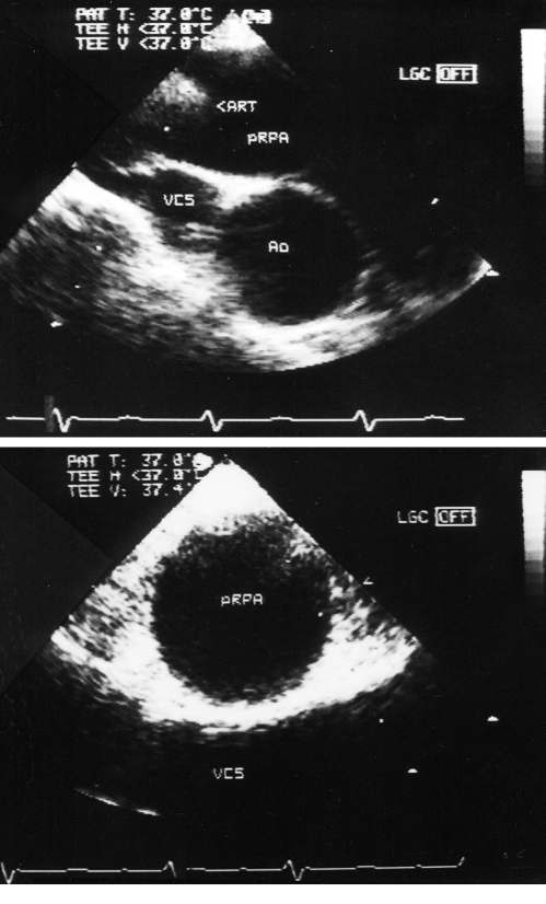
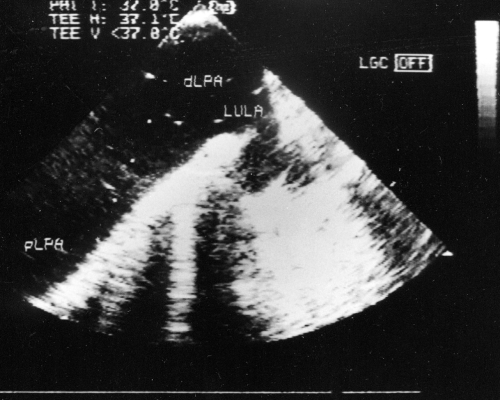
It is suggested that transesophageal echocardiography (TEE), by detecting thromboemboli in the proximal parts of the pulmonary arteries, is useful in the diagnosis of pulmonary embolism. However, the data on visualization of the pulmonary arteries are limited. The extent of the pulmonary arteries that can be precisely visualized during biplane TEE was assessed in 51 consecutive patients (23 female, 28 male, aged 56.6+/-12.5 years) without structural heart disease. The main pulmonary artery and the right pulmonary artery were detected in 96.1% and 94.1% of patients, respectively. Although the proximal part of the left pulmonary artery was found in only 47.0% of patients, its distal part was visualized in 92.2%. During TEE, proximal parts of the lobar arteries on both sides were visualized in 88.2% of patients. Thus, the central pulmonary arteries including proximal parts of the lobar branches can be precisely visualized by biplane TEE in the majority of patients. Only the proximal part of the left pulmonary artery is difficult to assess.
Keywords: Pulmonary arteries; Transesophageal echocardiography; Visualization.
Figures


References
-
- Pruszczyk P, Torbicki A, Kuch-Wocial A, Chlebus M, Miskiewicz ZC, Jedrusik P. Transoesophageal echocardiography for definitive diagnosis of haemodynamically significant pulmonary embolism. Eur Heart J. 1995;16:534–8. - PubMed
-
- Rittoo D, Sutherland GR, Samuel L, Flapan AD, Shaw TR. Role of transesophageal echocardiography in diagnosis and management of central pulmonary artery thromboembolism. Am J Cardiol. 1993;71:1115–8. - PubMed
-
- Wittlich N, Erbel R, Eichler A, et al. Detection of central pulmonary artery thromboemboli by transesophageal echocardiography in patients with severe pulmonary embolism. J Am Soc Echocardiogr. 1992;5:515–24. - PubMed
-
- Seward JB, Khandheria BK, Freeman WK, et al. Multiplane transesophageal echocardiography: image orientation, examination technique, anatomic correlations, and clinical applications. Mayo Clin Proc. 1993;68:523–51. - PubMed
-
- Fournier P, Augusseau-Richard MP, Charbonnier B, Pottier JM, Pigale C, Pacouret G. [Contribution of transesophageal echocardiography to the diagnosis of pulmonary embolism] Arch Mal Coeur Vaiss. 1994;87:459–65. - PubMed
LinkOut - more resources
Full Text Sources