. 2001 Summer;6(2):61-71.

Potent and use-dependent block of cardiac sodium channels by U-50,488H, a benzeneacetamide kappa opioid receptor agonist

Affiliations

Potent and use-dependent block of cardiac sodium channels by U-50,488H, a benzeneacetamide kappa opioid receptor agonist

M K Pugsley et al. Exp Clin Cardiol. 2001 Summer.

Abstract

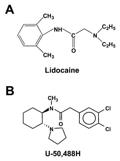
Objectives: To determine whether the kappa opioid receptor agonist U-50,488H, a benzacetamide derivative of the cyclo-hexane-1,2-diamine analgesics, may be a useful molecular probe to define the structural requirements of this class of drugs for cardiac sodium channel blockade.

Animals and methods: The electrophysiological effects of U-50,488H were compared with those of lidocaine, a clinically used class Ib antiarrhythmic agent, in rat heart sodium currents expressed in Xenopus laevis oocytes by using two-electrode voltage clamp.

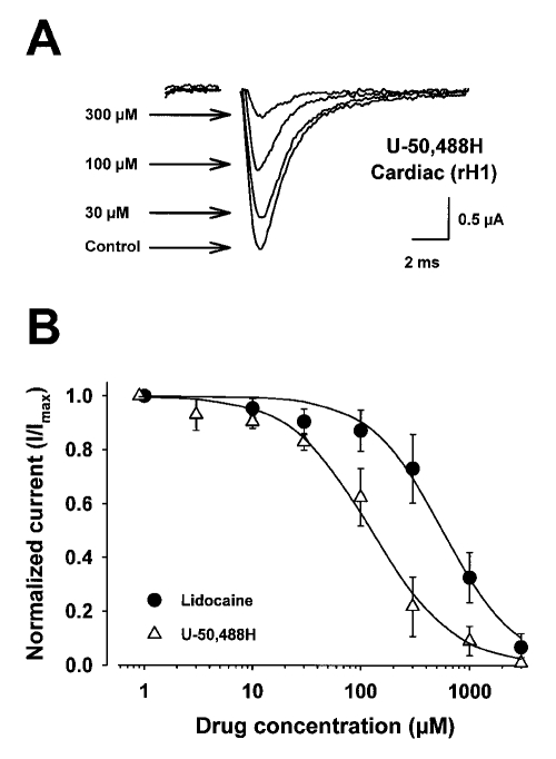
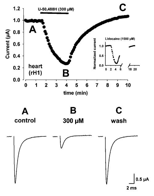
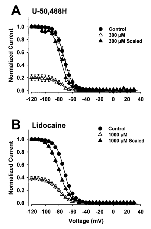
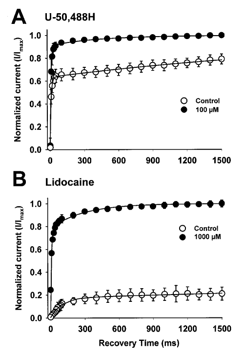
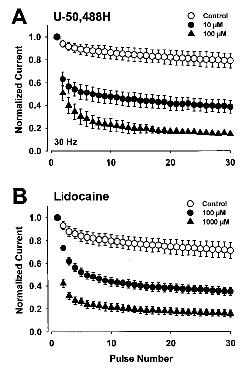
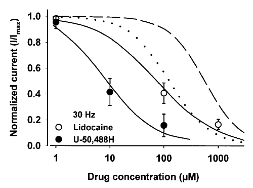
Results: Both U-50,488H and lidocaine produced a concentration-dependent tonic block of sodium current, but U-50,488H was approximately fourfold more potent than lidocaine. Both drugs produced a hyperpolarizing shift in the voltage dependence of sodium channel inactivation and both delayed recovery from inactivation. Both drugs exhibited use-dependent block, but U-50,488H showed a 1.8-fold increase in potency compared with lidocaine at a high frequency of stimulation (30 Hz).

Conclusions: The more potent tonic and use-dependent block of cardiac sodium channels by U-50,488H suggests that structural features of this molecule may provide it with a greater ability to block the channel. An understanding of these structural features may provide information needed in the development of novel arylacetamide-based antiarrhythmic drugs and insight into possible mechanisms describing channel block, resulting in a highly efficacious antiarrhythmic action in the heart.

Keywords: Antiarrhythmic agents; Membrane currents; Sodium channel; U-50,488H.


Figures

Figure 1
Figure 1
A Molecular structure of lidocaine (2-(diethylamino)-N-(2,6-dimethylphenyl)acetamide monohydrochloride). B General molecular structure of the arylacetamide compound U-50,488H (trans-(±)-3,4-dichloro-N-methyl-N-[7-(1-pyrrolidinyl)cyclohexyl]benzeneacetamide methane sulphonate)
Figure 2
Figure 2
Concentration-response curves for the effect of lidocaine and U-50,488H on rat heart (rH1) sodium currents expressed in Xenopus laevis oocytes. Currents were recorded by two-electrode whole-cell voltage clamp. Oocytes were injected with 50 ng of in vitro transcribed RNA encoding the rH1 sodium channel alpha-subunit. After two days of incubation at 20°C in ND-96 plus supplements, current-voltage curves were evoked by depolarizing the cell to various potentials from a fixed prepulse potential of −90 mV. The data were filtered at 3 kHz. Peak sodium currents, evoked every 6 s, were measured at test potentials that elicited maximum inward current (−10 mV). Peak sodium currents were measured again after 5 min perfusion of the cell at a flow rate of 1 to 2 mL/min of ND96 containing increasing concentrations of either U-50,488H or lido-caine. The data are shown as peak current normalized to the maximum current in the absence of drug (I/Imax). The curves described by the solid lines were fitted by the Hill equation where INa = [1 + ([A]/EC50)n]−1. INa describes the fraction of maximal current remaining after block by either drug, [A] is the concentration of drug, EC50 is the concentration of drug at half-maximal block, and n is the Hill coefficient describing the stoichiometry of the drug-channel interaction. Concentration-response curves are mean ± SD for five oocytes. The parameters of the fits are shown in Table 2
Figure 3
Figure 3
Time course for rat heart (rH1) sodium channel block by U-50,488H. Oocytes were held at −120 mV and currents were evoked by depolarizations to −10 mV every 6 s in the absence (A) and presence (B) of U-50,488H. At the time indicated by the bar, 300 μM U-50,488H was added to the solution perfusing the cell. Maximal rH1 sodium current amplitude is shown plotted against time. Current traces are shown under control conditions (A, control), at the time maximal block was attained with U-50,488H (B, 300 μM), and 5 min after removal of U-50,488H from the bath solution and recovery of pre-drug current amplitude (C, wash). The inset in the upper panel depicts the time course for 1000 μM lidocaine block of the rH1 sodium current under identical experimental conditions. Note the prolonged time scale required for current recovery from block
Figure 4
Figure 4
Effects of U-50,488H (300 μM) and lidocaine (1000 μM) on the voltage dependence of steady-state inactivation of rat heart sodium channels. Inactivation was examined using a two-pulse protocol in which oocytes were held at −120 mV and depolarized from −90 to +15 mV for 500 ms, followed by a test pulse to −5 mV for 22.5 ms to determine channel availability. The data points in both panels were determined from 5 oocytes and error bars represent SD. All curves were fitted with a two-state Boltzmann function. Inactivation curves are shown in the absence (control) and presence of either 300 μM U-50,488H or 1000 μM lidocaine for the heart channel. The data obtained in the presence of either drug are also scaled to the maximum current observed without drug to show the shifts in voltage-dependence
Figure 5
Figure 5
Effects of 100 μM U-50,488H (A) and 1000 μM lidocaine (B) on recovery from inactivation of rat heart sodium channels. Oocytes were held at −120 mV and depolarized with an inactivating pulse to −10 mV for 500 ms. This was followed by a variable recovery interval ranging from 10 to 1500 ms at −120 mV. The recovery interval was followed by a 20 ms test depolarization to −10 mV. The peak current amplitude elicited during the test pulse was normalized to the peak current amplitude during a 22 ms pulse elicited from a holding potential of −120 mV for the rat heart channel immediately before the recovery protocol. The mean ± SD for the normalized current is shown as a function of the recovery time for at least four oocytes either in the absence of drug or in the presence of 100 μM U-50,488H (A) or 1000 μM lidocaine (B). The smooth curves represent fits of the data to a double exponential equation
Figure 6
Figure 6
Effects of U-50,488H (A) and lidocaine (B) on use-dependent block of rat heart sodium channels in the absence (control) or presence of low or high concentrations of U-50,488H or lidocaine. A series of 20 ms depolarizing pulses to −10 mV was applied from a holding potential of −120 mV for rat heart channels at 30 Hz. The peak currents were normalized to the current during the first depolarization and plotted as a function of pulse number. The peak currents measured in the presence of drug were normalized to the current during the first depolarization in the presence of drug. Those currents measured approximately 10% of the current amplitudes in the absence of low concentrations of U-50,488H and lidocaine and approximately 40% in the absence of high concentrations of these drugs. This normalization emphasizes the amount of use-dependent block that developed independently of the tonic block by either drug
Figure 7
Figure 7
Concentration-response curves for the use-dependent block by U-50,488H and lidocaine on rat heart (rH1) sodium currents expressed in Xenopus laevis oocytes. Currents were recorded by two-electrode whole-cell voltage clamp. Oocytes were injected with in vitro transcribed RNA encoding the rH1 sodium channel alpha-subunit. The amount of use-dependent block is determined by the residual current that exists at the end of a 30 Hz series of 20 ms depolarizing pulses to −10 mV that were applied from a holding potential of −120 mV to rH1 channels compared with the current in the absence of drug. The data are shown as residual current normalized to the maximum current in the absence of drug (I/Imax). The smooth curves described by the solid lines were fitted by the Hill equation where INa = [1 + ([A]/EC50)n]−1. INa describes the fraction of maximal current remaining after block by either drug, [A] is the concentration of drug, EC50 is the concentration of drug at half-maximal block, and n is the Hill coefficient describing the stoichiometry of the drug-channel interaction. Concentration-response curves are mean ± SD for five oocytes. For comparison, the concentration-response curves describing tonic block (from Figure 2) of U-50,488H (smooth dotted curve) and lidocaine (short dashed curve) on rH1 sodium currents are shown when peak sodium currents were evoked every 6 s and measured at test potentials that elicited maximum inward current (−10 mV)

Similar articles

Cited by

References

    1. Goldin AL. Voltage-gated sodium channels. In: North RA, editor. Ligand- and Voltage-Gated Ion Channels. Boca Raton: CRC Press; 1995. pp. 73–112.
    1. Noda M, Ikeda T, Suzuki H, et al. Expression of functional Na+ channels from cloned cDNA. Nature. 1986;322:826–8. - PubMed
    1. Isom LL, DeJongh KS, Patton DE, et al. Primary structure and functional expression of the β1 subunit of the rat brain sodium channel. Science. 1992;256:839–42. - PubMed
    1. Goldin AL. Molecular analysis of Na+ channel inactivation. In: Perracjia C, editor. Hanbook of Membrane Channels, vol 1. San Diego: Academic Press; 1994. pp. 121–35.
    1. Hodgkin AL, Huxley AF. A quantitative description of membrane current and its application to conduction and excitation in nerve. J Physiol. 1952;117:500–44. - PMC - PubMed

LinkOut - more resources