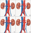
The clinical significance of a retroaortic left renal vein
- PMID: 20428432
- PMCID: PMC2858856
- DOI: 10.4111/kju.2010.51.4.276
The clinical significance of a retroaortic left renal vein
Abstract
Purpose: A retroaortic left renal vein (RLRV) is located between the aorta and the vertebra and drains into the inferior vena cava. Urological symptoms can be caused by increased pressure in the renal vein. To evaluate the clinical importance of RLRV, we reviewed patients' medical records and radiologic findings.
Materials and methods: Nine patients who were studied with multidetector computed tomography at our institution from January 2003 to December 2009 had urologic symptoms with RLRV. We retrospectively reviewed these patients' medical records and analyzed their clinical characteristics.
Results: The patients' mean age was 46.0+/-20.1 years (range, 17-65 years) and the male to female ratio was 5 to 4. The urologic symptoms of the initial diagnosis were various (hematuria: 5 of the 9 patients; left flank pain: 4 of the 9 patients; inguinal pain: 1 of the 5 male patients; and gross hematuria: 1 of the 9 patients). The distribution among the type I, II, III, and IV of RLRV was 6, 2, 1, and 0 patients, respectively. The concomitant diseases were ureteropelvic junction obstruction (UPJO; 2 of the 9 patients) and varicocele (2 of the 5 male patients). One patient with UPJO underwent pyeloplasty and the other patient with UPJO underwent nephrectomy due to a nonfunctional atrophied kidney. The microscopic hematuria was not resolved with conservative management for long-term follow-up.
Conclusions: Hematuria and inguinal or flank pain seem to be common in patients with RLRV. The most common type of RLRV was type I. It appeared that the microscopic hematuria continued in the long-term follow-up.
Keywords: Abnormalities; Renal veins; Tomography.
Conflict of interest statement
The authors have nothing to disclose.
Figures


References
-
- Mayo J, Gray R, St Louis E, Grosman H, McLoughlin M, Wise D. Anomalies of the inferior vena cava. AJR Am J Roentgenol. 1983;140:339–345. - PubMed
-
- Royal SA, Callen PW. CT evaluation of anomalies of the inferior vena cava and left renal vein. AJR Am J Roentgenol. 1979;132:759–763. - PubMed
-
- Hayashi M, Kume T, Nihira H. Abnormalities of renal venous system and unexplained renal hematuria. J Urol. 1980;124:12–16. - PubMed
-
- Thomas TV. Surgical implications of retroaortic left renal vein. Arch Surg. 1970;100:738–740. - PubMed
-
- Cuéllar i Calàbria H, Quiroga Gómez S, Sebastia Cerquedà C, Boyé de la Presa R, Miranda A, Alvarez-Castells A. Nutcracker or left renal vein compression phenomenon: multidetector computed tomography findings and clinical significance. Eur Radiol. 2005;15:1745–1751. - PubMed
LinkOut - more resources
Full Text Sources

