. 2010 Apr 13:4:15.
doi: 10.3389/fnana.2010.00015. eCollection 2010.

Reconstruction and dissection of the entire human visual pathway using diffusion tensor MRI

Affiliations

Reconstruction and dissection of the entire human visual pathway using diffusion tensor MRI

Sabine Hofer et al. Front Neuroanat. .

Abstract

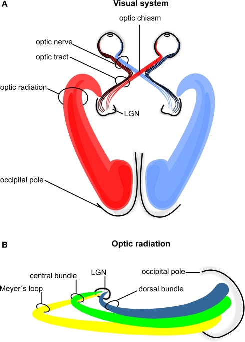
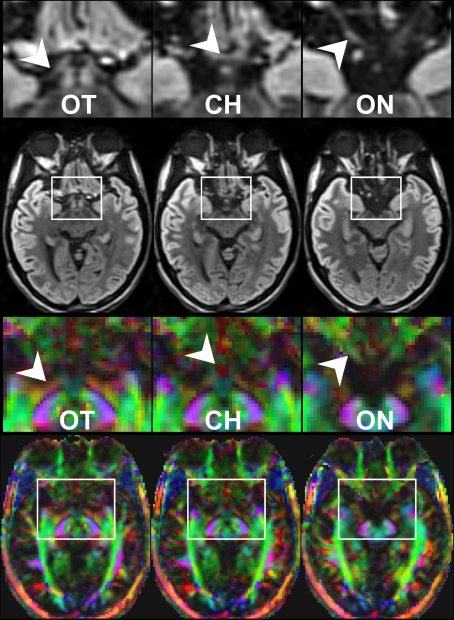
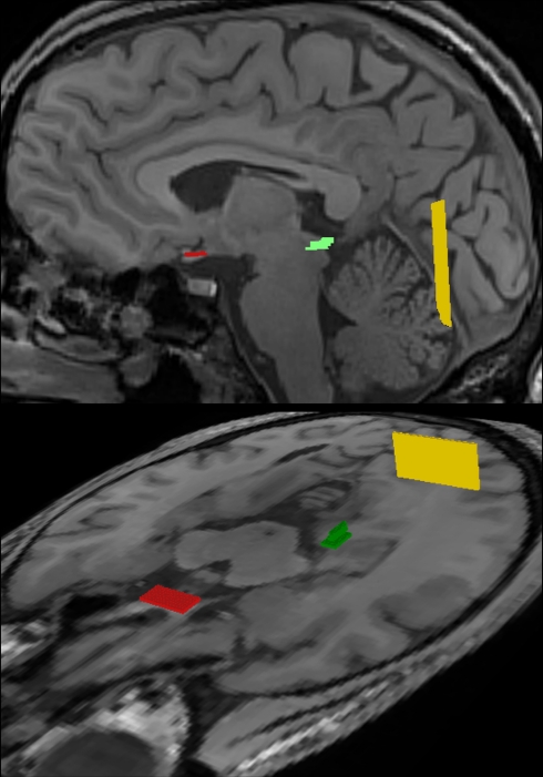
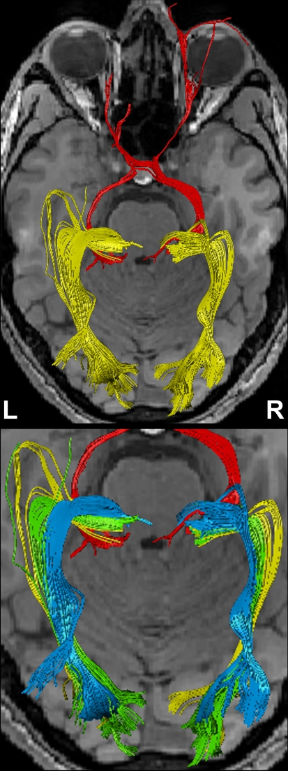
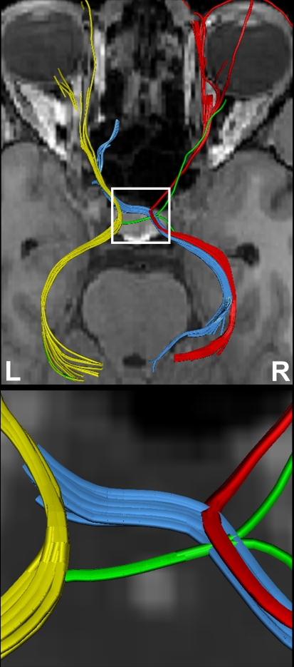
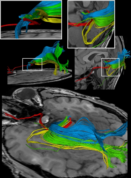
THE HUMAN VISUAL SYSTEM COMPRISES ELONGATED FIBER PATHWAYS THAT REPRESENT A SERIOUS CHALLENGE FOR DIFFUSION TENSOR IMAGING (DTI) AND FIBER TRACTOGRAPHY: while tracking of frontal fiber bundles may be compromised by the nearby presence of air-filled cavities, nerves, and eye muscles, the anatomic courses of the three main fiber bundles of the optic radiation are subject to pronounced inter-subject variability. Here, tractography of the entire visual pathway was achieved in six healthy subjects at high spatial accuracy, that is, at 1.8 mm isotropic spatial resolution, without susceptibility-induced distortions, and in direct correspondence to anatomic MRI structures. Using a newly developed diffusion-weighted single-shot STEAM MRI sequence, we were able to track the thin optic nerve including the nasal optic nerve fibers, which cross the optic chiasm, and to dissect the optic radiation into the anterior ventral bundle (Meyer's loop), the central bundle, and the dorsal bundle. Apart from scientific applications these results in single subjects promise advances in the planning of neurosurgical procedures to avoid unnecessary damage to the visual fiber system.

Keywords: DTI; Meyer's loop; fiber tractography; human brain; optic chiasm.


Figures

Figure 1
Figure 1
Schematic drawing of the human visual system. (A) Right- and left-hemispheric fiber pathways. (B) Left-hemispheric optic radiation comprising the anterior bundle termed Meyer's loop (yellow), the central bundle (green), and the dorsal bundle (blue). LGN, lateral geniculate nucleus.
Figure 2
Figure 2
Contiguous diffusion-weighted STEAM images (averaged across gradient directions) and color-coded FA maps covering the area of the optic nerve and chiasm without susceptibility artifacts at 1.8 mm isotropic spatial resolution (magnified views on top, arrowheads indicate respective areas). OT, optic tract; CH, chiasm; ON, optic nerve. Green = anterior–posterior, blue = superior–inferior, red = right–left.
Figure 3
Figure 3
Regions-of-interest (ROI) chosen for fiber tractography of the visual pathway in complementary views. The optic nerve and optic tract were delineated by combining a start ROI in the optic chiasm (red) with a target ROI in the LGN (green). Correspondingly, the optic radiation was obtained by a start ROI in the LGN (green) and a target ROI in the white matter next to primary visual areas (yellow). For further details see text.
Figure 4
Figure 4
(Top) Bi-hemispheric reconstructions of the optic nerve and tract (red) as well as of the optic radiation (yellow). (Bottom) Dissection of the optic radiation into Meyer's loop (yellow), central bundle (green), and dorsal bundle (blue). R, right, L, left.
Figure 5
Figure 5
(Top) Separation of right- and left-hemispheric fibers in the optic nerve and optic tract. (Bottom) Magnification of fiber tracks crossing the optic chiasm: tracks from the left eye to the left (yellow) and right LGN (blue), tracks from the right eye to the left (green) and right LGN (red). R, right, L, left.
Figure 6
Figure 6
Reconstructions of the optic nerve and tract (red), Meyer's loop (yellow), central bundle (green), and dorsal bundle (blue) in different views. (White boxes) Magnifications of the area around the LGN. (Upper left) Fiber bundles of the right hemisphere. (Upper right and bottom) Fiber bundles of the left hemisphere.

References

    1. Bürgel U., Schormann T., Schleicher A., Zilles K. (1999). Mapping of histologically identified long fiber tracts in human cerebral hemispheres to the MRI volume of a reference brain: position and spatial variability of the optic radiation. Neuroimage 10, 489–499 10.1006/nimg.1999.0497 - DOI - PubMed
    1. Coenen V. A., Huber K. K., Krings T., Weidemann J., Gilsbach J. M., Rohde V. (2005). Diffusion-weighted imaging-guided resection of intracerebral lesions involving the optic radiation. Neurosurg. Rev. 28, 188–195 10.1007/s10143-005-0385-6 - DOI - PubMed
    1. Dougherty R. F., Ben-Shachar M., Bammer R., Brewer A. A., Wandell B. A. (2005). Functional organization of human occipital-callosal fiber tracts. Proc. Natl. Acad. Sci. U.S.A. 102, 7350–7355 10.1073/pnas.0500003102 - DOI - PMC - PubMed
    1. Ebeling U., Reulen H. J. (1988). Neurosurgical topography of the optic radiation in the temporal lobe. Acta Neurochir. (Wien) 92, 29–36 10.1007/BF01401969 - DOI - PubMed
    1. Hofer S., Frahm J. (2006). Topography of the human corpus callosum revisited – comprehensive fiber tractography using diffusion tensor magnetic resonance imaging. Neuroimage 32, 989–994 10.1016/j.neuroimage.2006.05.044 - DOI - PubMed

LinkOut - more resources