

Folate-immunoglobulin G as an anticancer therapeutic antibody
- PMID: 20429546
- PMCID: PMC3812690
- DOI: 10.1021/bc900545h
Folate-immunoglobulin G as an anticancer therapeutic antibody
Abstract
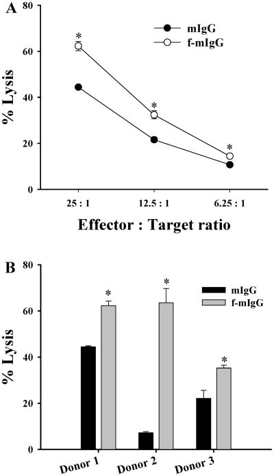
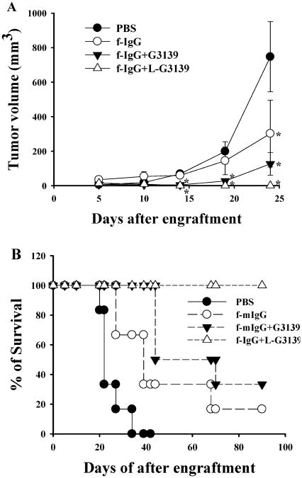
Folate receptor-alpha (FR) is a promising cellular marker for tumor-specific drug delivery. Conjugation of folic acid to therapeutic and imaging agents has been shown to enhance their delivery to FR (+) cancer cells in vitro and in tumor-bearing mice via an FR-mediated cellular uptake mechanism. In this study, immunoglobulin G (IgG) was conjugated to folate and evaluated as a therapeutic antibody against folate receptor (FR)-positive tumors. Murine IgG (mIgG) was conjugated to folate via an amide bond to yield folate-conjugated mIgG (f-mIgG) that contained an average of approximately 2.6 folates per molecule. Selective uptake of f-IgG by FR (+) tumor cells was determined by fluorescence microscopy and by flow cytometry. Lysis of L1210JF cells by NK cells from murine donors was increased 1.4-9.0-fold at the effector:target (E:T) ratio of 25:1, relative to control mIgG. In mice bearing L1210JF tumors, f-mIgG was found to significantly inhibit tumor growth and to have prolonged the median survival time (MeST). Significantly, the antitumor efficacy of f-mIgG was greatly increased when combined with liposomal G3139, an 18-mer phosphorothioate oligonucleotide. In fact, the combination resulted in a 100% cure rate among the tumor-bearing mice. Injection of f-mIgG significantly increased serum INF-gamma and IL-6 level in mice compared with mIgG and dramatically increased serum INF-gamma and IL-6 level when combined with liposomal G3139. These results suggested that f-IgG, a novel immunotherapy agent, has potent activity as a therapeutic antibody to the FR-positive cancer, and the therapeutic activity is enhanced by immunomodulatory agents.
Figures


Similar articles
-
In vivo antitumor activity of folate receptor-targeted liposomal daunorubicin in a murine leukemia model.Anticancer Res. 2005 Jan-Feb;25(1A):343-6. Anticancer Res. 2005. PMID: 15816557
-
Delivery of calf thymus DNA to tumor by folate receptor targeted cationic liposomes.Biomaterials. 2011 Sep;32(27):6614-20. doi: 10.1016/j.biomaterials.2011.05.037. Epub 2011 Jun 12. Biomaterials. 2011. PMID: 21665267 Free PMC article.
-
NK Cell-Mediated Antitumor Effects of a Folate-Conjugated Immunoglobulin Are Enhanced by Cytokines.Cancer Immunol Res. 2016 Apr;4(4):323-336. doi: 10.1158/2326-6066.CIR-15-0168. Epub 2016 Feb 10. Cancer Immunol Res. 2016. PMID: 26865456 Free PMC article.
-
Targeted drug delivery via folate receptors.Expert Opin Drug Deliv. 2008 Mar;5(3):309-19. doi: 10.1517/17425247.5.3.309. Expert Opin Drug Deliv. 2008. PMID: 18318652 Review.
-
Folate receptor-mediated drug targeting: from therapeutics to diagnostics.J Pharm Sci. 2005 Oct;94(10):2135-46. doi: 10.1002/jps.20457. J Pharm Sci. 2005. PMID: 16136558 Review.
Cited by
-
Induction of ADCC by a folic acid-mAb conjugate prepared by tryptophan-selective reaction toward folate-receptor-positive cancer cells.RSC Adv. 2020 Apr 29;10(28):16727-16731. doi: 10.1039/d0ra03291c. eCollection 2020 Apr 23. RSC Adv. 2020. PMID: 35498849 Free PMC article.
-
Fc-Small Molecule Antibody Mimetics.Bioconjug Chem. 2015 Dec 16;26(12):2311-4. doi: 10.1021/acs.bioconjchem.5b00530. Epub 2015 Nov 13. Bioconjug Chem. 2015. PMID: 26536496 Free PMC article.
-
IgA Fc-folate conjugate activates and recruits neutrophils to directly target triple-negative breast cancer cells.Breast Cancer Res Treat. 2018 Dec;172(3):551-560. doi: 10.1007/s10549-018-4941-5. Epub 2018 Aug 28. Breast Cancer Res Treat. 2018. PMID: 30155754 Free PMC article.
-
Epigenetic Regulation and Dietary Control of Triple Negative Breast Cancer.Front Nutr. 2020 Sep 8;7:159. doi: 10.3389/fnut.2020.00159. eCollection 2020. Front Nutr. 2020. PMID: 33015128 Free PMC article. Review.
-
Nucleic acid-based nanoengineering: novel structures for biomedical applications.Interface Focus. 2011 Oct 6;1(5):702-24. doi: 10.1098/rsfs.2011.0040. Epub 2011 Jun 28. Interface Focus. 2011. PMID: 23050076 Free PMC article.
References
-
- Toffoli G, Cernigoi C, Russo A, Gallo A, Bagnoli M, Boiocchi M. Overexpression of folate binding protein in ovarian cancers. Int J Cancer. 1997;74:193–198. - PubMed
-
- Bueno R, Appasani K, Mercer H, Lester S, Sugarbaker D. The alpha folate receptor is highly activated in malignant pleural mesothelioma. J Thorac Cardiovasc Surg. 2001;121:225–233. - PubMed
-
- Parker N, Turk E, Westrick MJ, Lewis JD, Low PS, Leamon CP. Folate receptor expression in carcinomas and normal tissues determined by a quantitative radioligand binding assay. Anal Biochem. 2005;338:284–293. - PubMed
-
- Ross JF, Chaudhuri PK, Ratnam M. Differential regulation of folate receptor isoforms in normal and malignant tissues in vivo and in established cell lines. Physiologic and clinical implications Cancer. 1994;73:2432–2443. - PubMed
-
- Weitman SD, Frazier KM, Kamen BA. The folate receptor in central nervous system malignancies of childhood. J Neurooncol. 1994;21:107–112. - PubMed
MeSH terms
Substances
Grants and funding
LinkOut - more resources
Full Text Sources
Other Literature Sources
Medical
Miscellaneous
