

Eosinophilic pneumonia associated with bleomycin in a patient with mediastinal seminoma: a case report
- PMID: 20429899
- PMCID: PMC2868877
- DOI: 10.1186/1752-1947-4-126
Eosinophilic pneumonia associated with bleomycin in a patient with mediastinal seminoma: a case report
Abstract
Introduction: Lung toxicities resulting from the chemotherapeutic agent bleomycin encompass a variety of pathological changes, including bronchiolitis obliterans organizing pneumonia, interstitial pneumonitis and progressive interstitial fibrosis. We report a rare case of eosinophilic pneumonia associated with bleomycin.
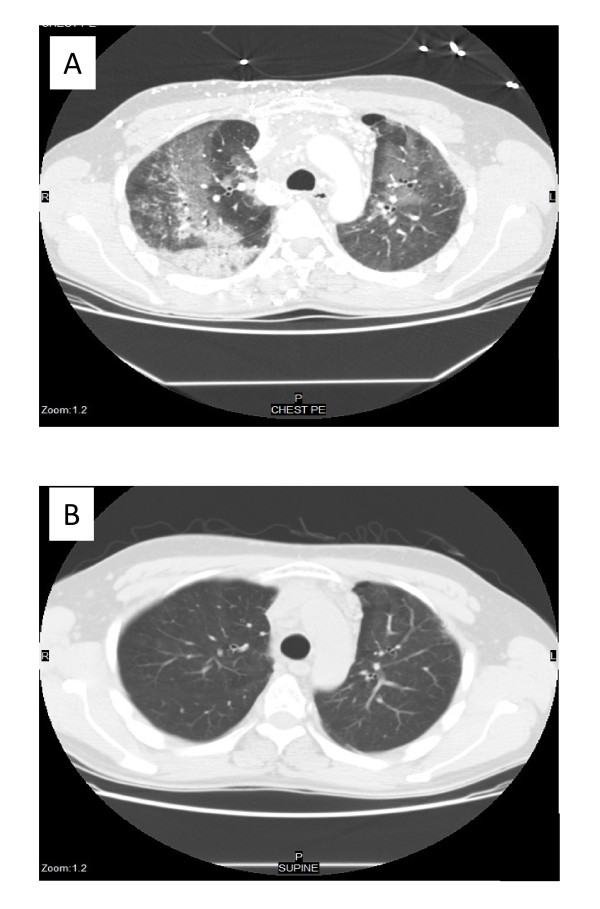
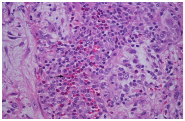
Case presentation: A 44-year-old Hispanic man with a primary mediastinal seminoma complicated by superior vena cava syndrome underwent treatment with four cycles of bleomycin, etoposide and cisplatin. He had a complete positive response to the chemotherapy. However, three months after treatment he presented with shortness of breath and severe hypoxemia associated with peripheral eosinophilia. Computed tomography showed bilateral diffuse interstitial infiltrates that were refractory to antibiotic treatment. A lung biopsy showed eosinophilic pneumonia. He was subsequently treated with high-dose prednisone, resulting in a complete resolution of his symptoms and lung infiltrates.
Conclusion: This case illustrates that eosinophilic pneumonia may be a late sequela of bleomycin toxicity, and may respond dramatically to steroid treatment.
Figures
References
-
- Bleomycin toxicities. http://www.pneumotox.com
-
- Cooper JA Jr, White DA, Matthay RA. Drug-induced pulmonary disease. Part 1: Cytotoxic drugs. Am Rev Respir Dis. 1986;133(2):321–340. - PubMed
-
- Ginsberg SJ, Comis RL. The pulmonary toxicity of antineoplastic agents. Semin Oncol. 1982;9(1):34–51. - PubMed
LinkOut - more resources
Full Text Sources
