. 2010 Aug;267(1-2):119-36.
doi: 10.1016/j.heares.2010.04.003. Epub 2010 Apr 27.

Areas of cat auditory cortex as defined by neurofilament proteins expressing SMI-32

Affiliations

Areas of cat auditory cortex as defined by neurofilament proteins expressing SMI-32

Jeffrey G Mellott et al. Hear Res. 2010 Aug.

Abstract

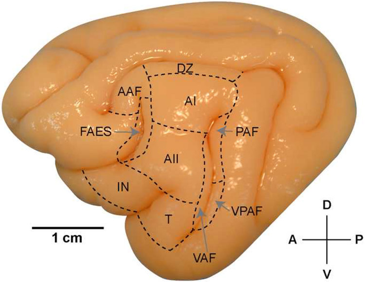
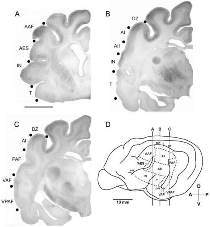
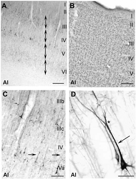
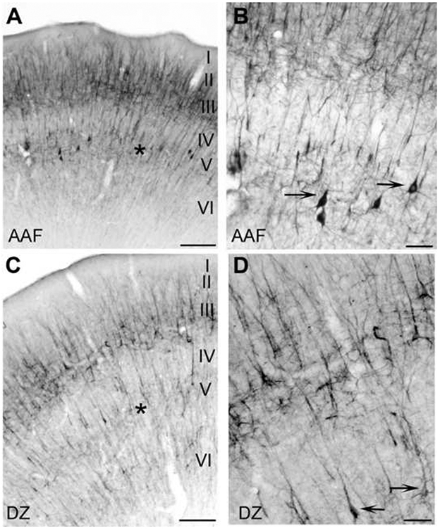
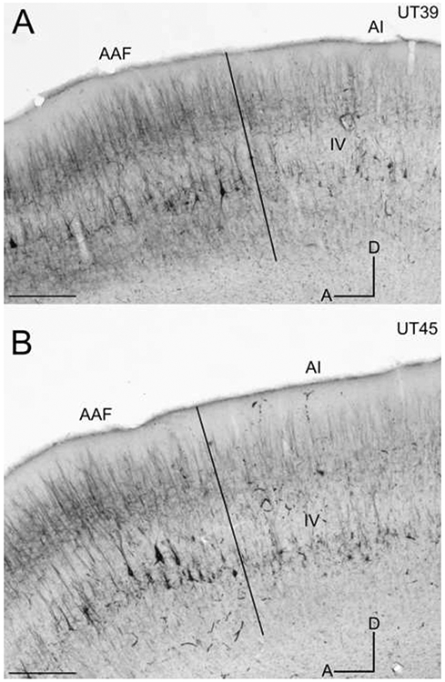
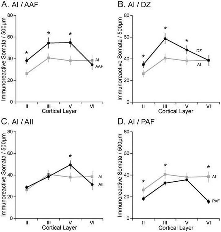
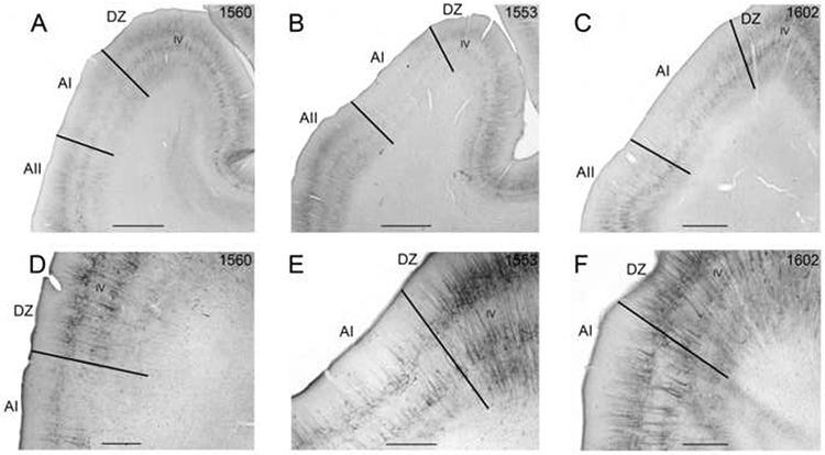
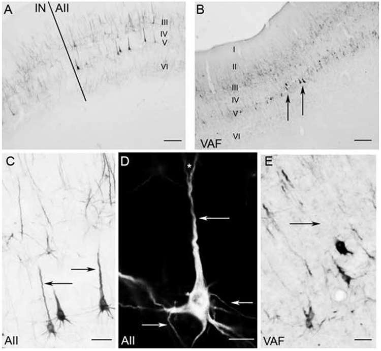
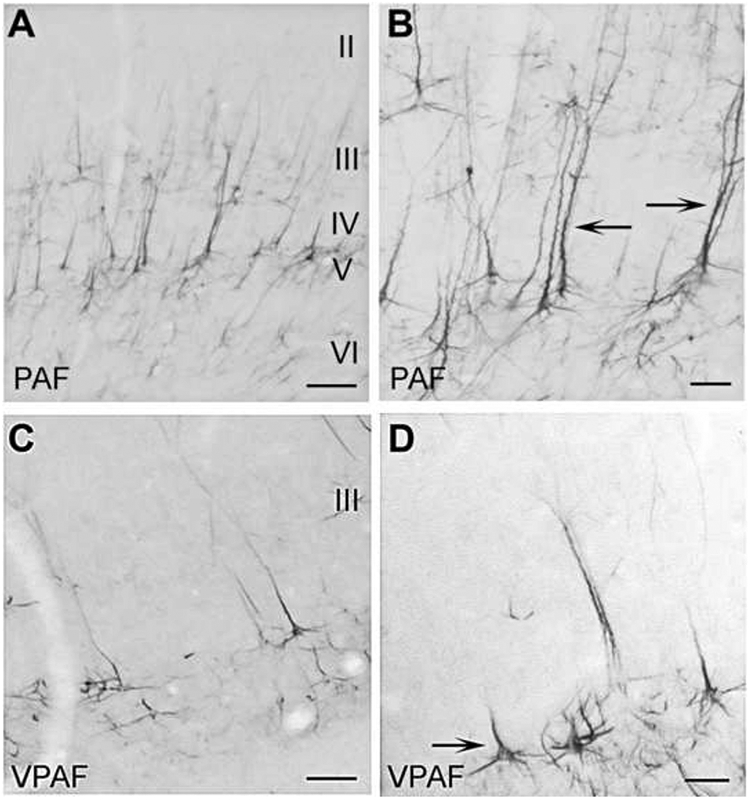
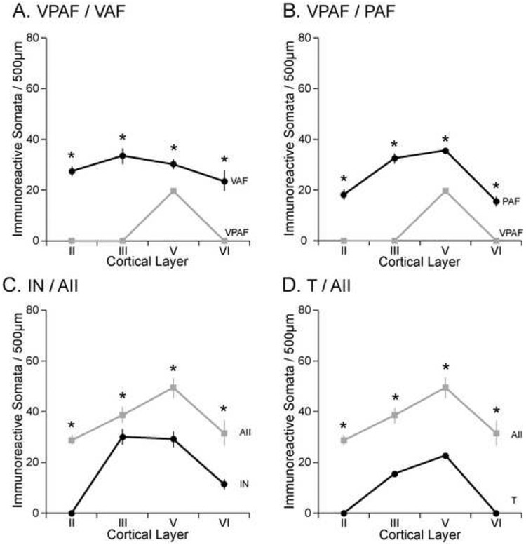
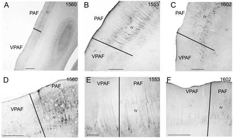
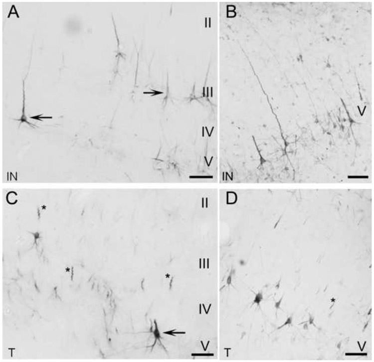
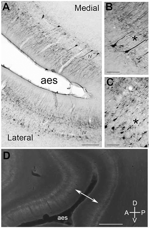
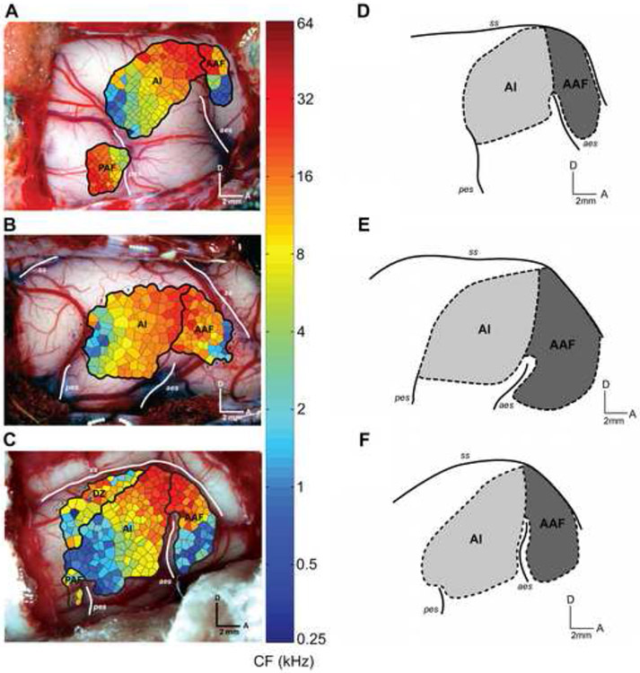
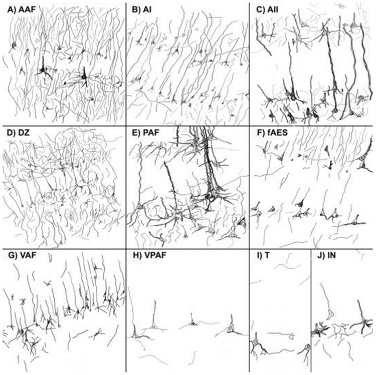
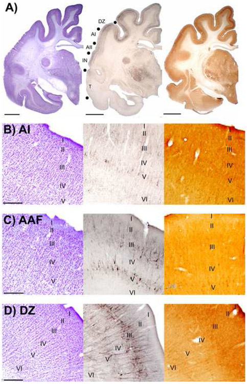
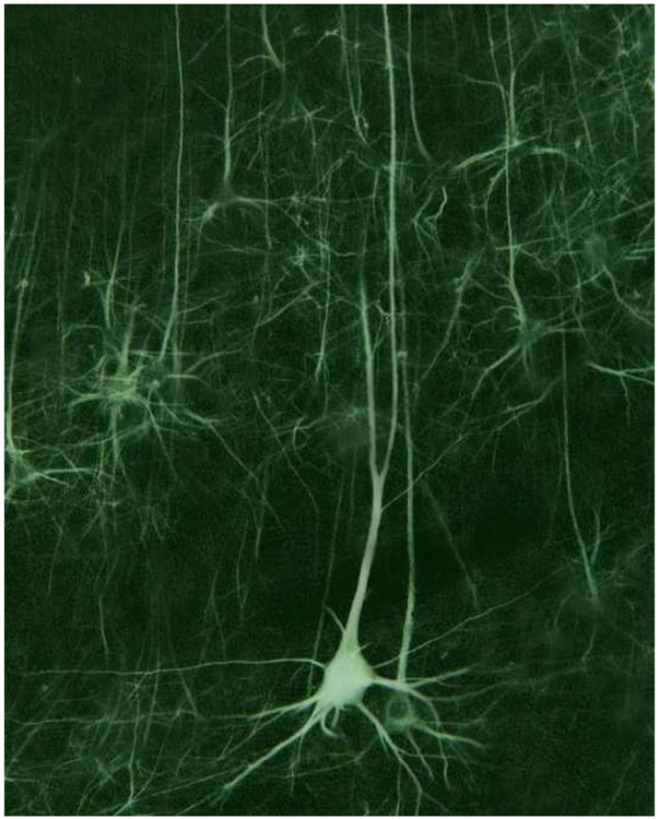
The monoclonal antibody SMI-32 was used to characterize and distinguish individual areas of cat auditory cortex. SMI-32 labels non-phosphorylated epitopes on the high- and medium-molecular weight subunits of neurofilament proteins in cortical pyramidal cells and dendritic trees with the most robust immunoreactivity in layers III and V. Auditory areas with unique patterns of immunoreactivity included: primary auditory cortex (AI), second auditory cortex (AII), dorsal zone (DZ), posterior auditory field (PAF), ventral posterior auditory field (VPAF), ventral auditory field (VAF), temporal cortex (T), insular cortex (IN), anterior auditory field (AAF), and the auditory field of the anterior ectosylvian sulcus (fAES). Unique patterns of labeling intensity, soma shape, soma size, layers of immunoreactivity, laminar distribution of dendritic arbors, and labeled cell density were identified. Features that were consistent in all areas included: layers I and IV neurons are immunonegative; nearly all immunoreactive cells are pyramidal; and immunoreactive neurons are always present in layer V. To quantify the results, the numbers of labeled cells and dendrites, as well as cell diameter, were collected and used as tools for identifying and differentiating areas. Quantification of the labeling patterns also established profiles for ten auditory areas/layers and their degree of immunoreactivity. Areal borders delineated by SMI-32 were highly correlated with tonotopically-defined areal boundaries. Overall, SMI-32 immunoreactivity can delineate ten areas of cat auditory cortex and demarcate topographic borders. The ability to distinguish auditory areas with SMI-32 is valuable for the identification of auditory cerebral areas in electrophysiological, anatomical, and/or behavioral investigations.


Figures

Figure 1.
Figure 1.
Lateral view of the left hemisphere of the cat brain. The ten auditory cortical areas identified are labeled. For abbreviations, see List. Scale bar=10mm.
Figure 2.
Figure 2.
Macroscopic views of coronal sections showing SMI-32 immunoreactivity. Even at low magnification the areal boundaries between neighboring regions and differing intensities can be identified (filled black circles). A: The anterior-most cortical regions identified (AAF, fAES, IN) and surrounding area (T). Dorsal regions were more heavily reactive, specifically within layer III, than the ventral regions, IN and T. Scale bar = 1cm and applies to A, B, & C. B: Middle ectosylvian regions (AI, AII, T) and neighboring regions (DZ, IN). C: Posterior ectosylvian regions (PAF, VPAF) along with middle ectosylvian VAF and neighboring regions (DZ, AI). D: Lateral view of the left cerebrum identifying the coronal positions of sections A, B and C. For abbreviations, see List.
Figure 3.
Figure 3.
Neurofilament protein expressed within AI depicting characteristic properties of the SMI-32 labeling. A: Reactive profile of each layer spanning AI. Note that most reactivity lies within layers III and V. Layers I and IV are barren of SMI-32 expression, which is consistent throughout each of the other nine auditory cortical areas. Double-headed arrows show that through SMI-32 application, laminar organization can be identified. B: Adjacent section to (A) reacted for the presence of Nissl. C: Photomicrograph at x100 revealing characteristics of reacted somata and dendrites. Arrows indicate apical dendrites that arose from layer V somata. Most apical dendrites and their corresponding somata were immunoreactive. D: A layer V pyramidal cell intensely immunoreactive to the SMI-32 antibody at x600. The arrow shows the well reacted apical dendrite. The asterisk indicates dendritic bifurcation. Well labeled bifurcations of layer V pyramidal calls are a prominent feature of AI. Scale bars: A and B=300μm; C=200μm; D=40μm.
Figure 4.
Figure 4.
Expression profiles of dorsal regions AAF (A, B) and DZ (C, D) at x40 (A, C) and x100 (B, D). A: SMI-32 reactivity across all cortical layers of AAF. AAF is characterized by an intense reaction, particularly layers II, III and V. Asterisk indicates layer V. B: Higher magnification (x100) of SMI-32 expression in AAF. Arrows indicate layer V somata. Robust labeling of large (>30μm) pyramidal cells throughout layer V is a unique characteristic of AAF. C: Photomicrograph of SMI-32 expression throughout all cortical layers of DZ. A distinguishing feature of DZ is the strong reactivity throughout layers II and III. Note that layer V in AAF was more intensely labeled than layer V of DZ (asterisks). D: Higher magnification (x100) of SMI-32 expression in DZ. Arrows indicate layer V somata reactive to SMI-32. Scale bars: A and C =400μm; B and D=100μm.
Figure 5.
Figure 5.
SMI-32 immunoreactivity in sagittal sections through the middle ectosylvian gyrus. Layer IV is labeled in each and borders are marked by black lines. A: The AI/AAF border at x200 in case UT39. B: The AI/AAF border at x200 in case UT45. D=dorsal; A=anterior. Scale bars: A&B = 1000μm.
Figure 6.
Figure 6.
Comparison of somata immunoreactivity between AI and each of its neighbors. A: AAF, B: DZ, C: AII, D: PAF. Each border shown is transitional. Graphs show mean ± SE immunoreactivity in each of the four immunoreactive layers (II, III, V, VI). Asterisks indicate significant difference at p<0.01.
Figure 7.
Figure 7.
SMI-32 immunoreactivity in coronal sections through AI of three cases. Layer IV is labeled in each and borders are marked by black lines. A-C: Low magnification photomicrographs of AI in cases 1560, 1553, and 1602. D: The AI/DZ border at x200 in case 1560. E: The AI/DZ border at x400 in case 1553. F: The AI/DZ border at x400 in case 1602. Scale bars: A-C = 2000μm; D-F = 1000μm.
Figure 8.
Figure 8.
SMI-32 reactivity in the middle ectosylvian areas AII (A,C,D) and VAF (B,E) A: Border of AII and neighboring cortical region IN. B: Low magnification photomicrograph of SMI-32 expression in VAF. Note heavy reactivity in layer V somata (arrows). C: Magnification (x100) of AII layer V somata. Arrows indicate the characteristic well labeled apical dendrites. D: A layer V pyramidal cell in AII at x600 under darkfield illumination. Arrows indicate the well defined dendrites that were commonly identified. The asterisk indicates a commonly observed bifurcation in an apical dendrite. E: Layer V somata in VAF at x100. The arrow shows the lack of a well-labeled apical dendrite, which is commonly absent from layer V somata in VAF. Scale bars: A and B=500μm; C and E=50μm; D=40μm.
Figure 9.
Figure 9.
SMI-32 expression in the posterior ectosylvian regions PAF (A,B) and VPAF (C,D). A: PAF has a moderate expression of SMI-32 throughout the supra- and infragranular layers. B: Many labeled pyramidal cells in layer V also have highly reactive apical dendrites (arrows). C: In contrast to PAF, VPAF has weak to no reactivity evident in the supragranular layers. D: Smaller, truncated somata are characteristic of VPAF layer V. Scale bars: A and C =300μm; B and D =40μm.
Figure 10.
Figure 10.
Comparison of somata immunoreactivity between areas that form “clear-cut” borders. A: VPAF/VAF, B: VPAF/PAF, C: IN/AII, D: T/AII. Graphs show mean ± SE immunoreactivity in each of the four immunoreactive layers. Asterisks indicate significant difference at p<0.01.
Figure 11.
Figure 11.
SMI-32 immunoreactivity in coronal sections through the posterior ectosylvian gyrus. Layer IV is labeled in each and borders are marked by black lines. A-C: Low magnification photomicrographs of the PAF/VPAF border in cases 1560, 1553, and 1602. D: The PAF/VPAF border at x200 in case 1560. E: The PAF/VPAF border at x400 in case 1553. F: The PAF/VPAF border at x200 in case 1602. Scale bars: A = 2000μm; B-D&F = 1000μm; E = 500μm.
Figure 12.
Figure 12.
SMI-32 expression profiles in limbic areas IN (A,B) and T (C,D). A,C are coronal sections and B,D are sagittal sections. A,B: Moderate to weak immunoreactivity spans across area IN, although somata and dendrites are readily visible and defined. In area IN, somata generally have a smaller stature than the more dorsal auditory regions. C,D: Layer V of area T is the most reactive layer. Traces of reactivity can be identified in layer III, but layers II and VI are barren. The arrow points to a layer V pyramidal cell in area T. Asterisks identify commonly found dendritic fragments strewn throughout area T. Scale bars = 50μm.
Figure 13.
Figure 13.
SMI-32 expression in the anterior ectosylvian (AE) sulcus. A: The photomicrograph shows possible SMI-32 subregions within the AE sulcus, medial and lateral. B: At higher power, a characteristic of the medial portion of the AE sulcus is the more full and reactive apical dendrites (*). C: The lateral portion of the AE sulcus has a more fragmented SMI-32 reactivity throughout layer V somata. D: Expression profile of the AE sulcus from another brain prepared sagittally. The double-headed arrow points to reactive disparity evident between supragranular layers of the anterior and posterior regions of the AE sulcus. Scale bars: A = 1000μm; B&C = 50μm; D = 2000μm.
Figure 14.
Figure 14.
The right hemisphere of three brains showing the A1-AAF border as defined by tonotopy (A-C) and the A1-AAF border as defined by SMI-32 reactivity (D-F). A-C) Characteristic frequency representation of two auditory cortical fields. Border was defined based on tonotopic organization. Each polygon represents an estimation of the cortical area with similar response properties as the recording site. The color of each polygon identifies the characteristic frequency at the recorded cortical location. D-F) Location of the A1-AAF border as defined by SMI-32 staining. A1 is shown in light grey and AAF is shown in dark gray. Note the similarities in the position of the border as defined by each method. Sulci are highlighted with thick white lines and indicated by italics: pes, posterior ectosylvian sulcus; ss, suprasylvian sulcus; aes, anterior ectosylvian sulcus. D, dorsal; A, anterior. Scale: 2 mm.
Figure 15.
Figure 15.
Illustrations of each cortical area examined for SMI-32 immunoreactivity spanning through layers III-V. A: AAF at x200 magnification. Intense immunoreactivity is common throughout AAF. B: AI at x200 magnification. A moderate and even reactivity in layers III and V is a key feature of AI. C: AII at x400 magnification. AII is readily identified by the robust, well-defined and intense labeling of its layer V pyramidal cells. D: DZ at x200 magnification. Similar to AAF, DZ is characterized by its heavily reactive supragranular layers. E: PAF at x600 magnification. Moderate reactivity with patches of larger well labeled layer V somata is characteristic of PAF. F: fAES at x600. fAES displayed moderate reactivity among layers III and V and the size of cells and intensity appear to vary as the sulcus’ conformation changes. G: VAF at x200 magnification. While VAF’s expression profile doesn't particularly stand out, its moderate neuropil immunostaining in layer V is striking enough to discern the region when compared to ventral neighbors VPAF and T. H: VPAF at x600 magnification. Barren supragranular layers and sparsely reactive layer V identify this area as the least immunoreactive of the ten examined regions. I: T at x600 magnification. This ventral region is lightly-moderately labeled with little intensity. Also, smaller cell diameters are more prevalent in T than the dorsal regions. J: IN at x600 magnification. The ventral region IN displayed a light to moderate label with smaller cell bodies that were reactive in layer V.
Figure 16.
Figure 16.
Areal comparisons of sections reacted for the presence of Nissl bodies, SMI-32 reactivity, and cytochrome oxidase (CO) reactivity in auditory cortex. A: Left hemisphere macroscopic views of adjacent coronal sections reacted for Nissl, SMI-32, and CO, respectively. Representative auditory areas are labeled on the middle panel. Borders between areas are indicated with filled black circles. Scale bars = 1cm. B: Adjacent coronal sections through AI reacted for Nissl, SMI-32, and CO, respectively. Photomicrographs were taken at x40 and applies to B, C, and D. Scale bar = 500μm and applies to B, C, and D. C: Adjacent coronal sections through AAF reacted for Nissl, SMI-32, and CO, respectively. D: Adjacent coronal sections through DZ reacted for Nissl, SMI-32, and CO, respectively.
Proposed Cover Illustration
Darkfield photomicrograph of pyramidal cells immunoreactive for SMI-32 in layers III and V of the posterior auditory field of the cat (magnification =200x).

Similar articles

Cited by

References

    1. Ashwell KWS, Zhang L-L, Marotte LR, 2005. Cyto-and chemoarchitecture of the cortex of the tammar wallaby (macropus eugenii): areal organization. Brain Behav. Evol 66, 114–136. - PubMed
    1. Baldauf ZB, 2005. SMI-32 parcellates the visual cortical areas of the marmoset. Neurosci. Lett 383, 1–2. - PubMed
    1. Beaver BV, Reed W, Leary S, McKiernan B, Bain F, Schultz R, Bennett BT, Pascoe P, Schull E, Cork LC, Francis-Floyd R, Amass KD, Johnson RJ, Schmidt RH, Underwood W, Thorton GW, Kohn B, 2001. 2000 Report of the American Veterinary Medical Association Panel of Euthanasia. J. Am. Vet. Med. Assoc 218, 669–696. - PubMed
    1. Beneyto M, Winer JA, Larue DT, Prieto JJ, 1998. Auditory connections and neurochemistry of the sagulum. J. Comp. Neurol 401, 329–351. - PubMed
    1. Bickford MA, Guido W, Godwin DW, 1998. Neurofilament proteins in Y-cells of the cat lateral geniculate nucleus: normal expression and alteration with visual deprivation. J. Neurosci 18, 6549–6557. - PMC - PubMed

Publication types

Substances