Review
. 2010 Mar;29(1):3-9.
doi: 10.1016/j.sder.2010.03.001.

New insights in the immunologic basis of psoriasis

Affiliations
Review

New insights in the immunologic basis of psoriasis

Kristine E Nograles et al. Semin Cutan Med Surg. 2010 Mar.

Abstract

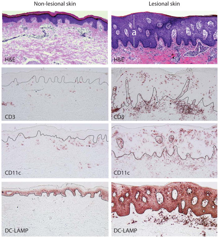
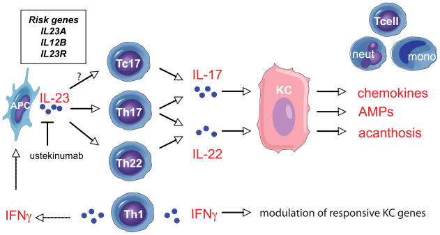
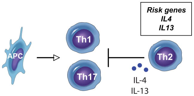
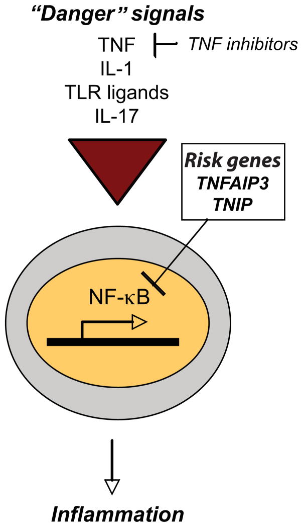
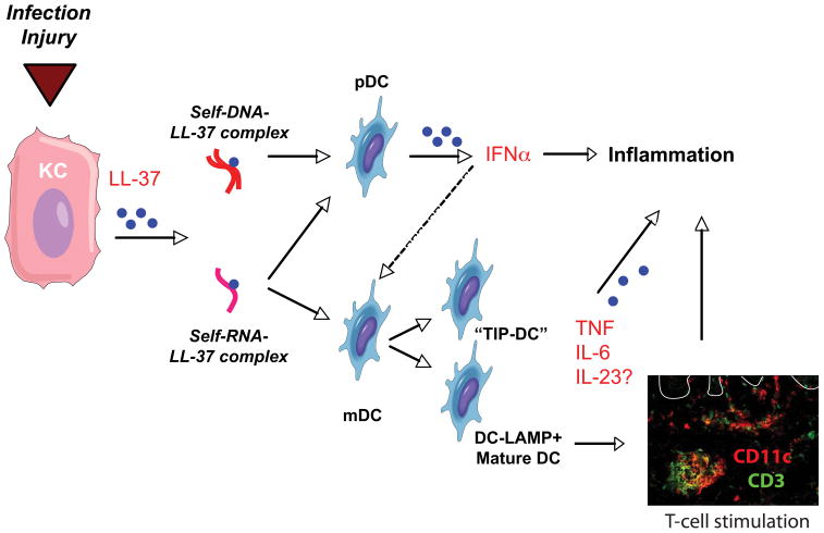
Psoriasis vulgaris is a multifactorial heritable disease characterized by severe inflammation resulting in poorly differentiated, hyperproliferative keratinocytes. Recent advances in genetic analyses have implicated components regulating the interleukin (IL)-23 and nuclear factor-kappaB pathways as risk factors for psoriasis. These inflammatory pathways exhibit increased activity in skin lesions, and promote secretion of various cytokines, such as IL-17 and IL-22. Unrestrained, the activated inflammatory cytokine network in psoriasis may trigger a vicious cycle of inflammation and cellular proliferation that ultimately results in lesion formation. These advances in genetic analyses, together with the progress made in targeted biological therapy, pave the path to tailor treatment on the basis of an individual's genetic and immunologic profile.


Conflict of interest statement

Disclosure: No relevant conflicts of interest in relations to this article.

Figures

Figure 1
Figure 1
Comparative histologic pictures of non-lesional and lesional psoriatic skin demonstrate marked acanthosis (a) and dermal inflammation (i) in psoriasis lesions compared to non-lesional skin (H&E stain). Inflammatory infiltrates in the psoriatic lesion consist of numerous T cells (CD3) as well as dendritic cells (CD11c), many of which are mature (DC-LAMP).
Figure 2
Figure 2. Model of immune interactions in the psoriatic lesion
Antigen-presenting cells (APC) produce IL-23 and stimulate Th17 and Th22 cells, and possibly Tc17 cells, to release IL-17 and IL-22. Keratinocytes (KC), in response to IL-17, upregulate pro-inflammatory chemokines that attract T cells, neutrophils (neut) and mononuclear cells (mono) into the lesion. IL-22 promotes epidermal acanthosis, while both cytokines trigger anti-microbial protein (AMP) production. IFNγ, from Th1 cells, modulates numerous KC responsive genes, and stimulates APCs to release IL-23. Ustekinumab, an FDA-approved monoclonal antibody, blocks the p40 sub-unit of IL-23. Recently identified genes associated with psoriasis (box) include IL23A, IL12B and IL23R.
Figure 3
Figure 3. Model of Th1-Th2-Th17 interactions
Effector T cells subsets stimulated by antigen-presenting cells (APC) in psoriasis include Th1 and Th17 cells. Th2 cells and associated cytokines, IL-4 and IL-13, that can suppress Th1 and Th17 activity, are decreased in psoriasis. Genes that confer risk of having psoriasis include IL4 and IL13 (box).
Figure 4
Figure 4. NFκB pathway in psoriasis
Multiple “danger” signals, including TNF, IL-1, toll-like receptor (TLR) ligands and IL-17, may stimulate the transcription factor, nuclear factor kappa-light-chain-enhancer of activated B cells (NFκB), to translocate into the nucleus and promote the transcription of inflammatory genes. Gene polymorphisms that promote unregulated NFkB activity may contribute to psoriasis susceptibility. Genome-wide associated studies (GWAS) have identified polymorphisms in TNFAIP3 and TNIP1, both negative regulators of the NFκB pathway (box).
Figure 5
Figure 5. Potential initiators of the inflammatory cascade in psoriasis
Infection and injury stimulate keratinocytes (KC) to release the anti-microbial, cathelicidin (LL-37). LL-37 forms complexes with self-DNA from damaged cells, and stimulates plasmacytoid dendritic cells (pDC) to release IFNα that activates myeloid dendritic cells (mDC). Simultaneously, LL-37 might form complexes with self-RNA to stimulate pDCs, as well as mDCs triggering the release of inflammatory cytokines TNF, IL-6 and, possibly, IL-23. Activation of mDCs by self-RNA-LL37 complexes promotes maturation of dendritic cells (DC-LAMP+ mature DCs) that enhances antigen-presenting capabilities to T cells. Double-label immunofluorescence demonstrates proximity of dendritic cells (CD11c, red) and T cells (CD3, green) in psoriatic dermis. White line delineates dermo-epidermal junction.

References

    1. Christophers E. Psoriasis--epidemiology and clinical spectrum. Clin Exp Dermatol. 2001;26(4):314–320. - PubMed
    1. Rongioletti F, Fiorucci C, Parodi A. Psoriasis induced or aggravated by drugs. J Rheumatol Suppl. 2009;83:59–61. - PubMed
    1. Gudjonsson J, Elder J. Psoriasis. In: Wolff K, Goldsmith L, Katz S, et al., editors. Fitzpatrick’s Dermatology in General Medicine. New York: McGraw-Hill; 2007.
    1. Lowes MA, Bowcock AM, Krueger JG. Pathogenesis and therapy of psoriasis. Nature. 2007;445(7130):866–873. - PubMed
    1. Ellis CN, Gorsulowsky DC, Hamilton TA, et al. Cyclosporine improves psoriasis in a double-blind study. JAMA. 1986;256(22):3110–3116. - PubMed

Publication types