. 2010 May;137(10):1721-9.
doi: 10.1242/dev.044586.

Hedgehog signaling controls mesenchymal growth in the developing mammalian digestive tract

Affiliations

Hedgehog signaling controls mesenchymal growth in the developing mammalian digestive tract

Junhao Mao et al. Development. 2010 May.

Abstract

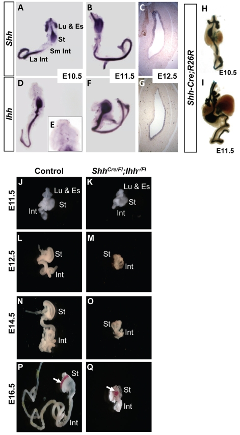
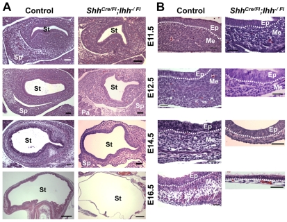
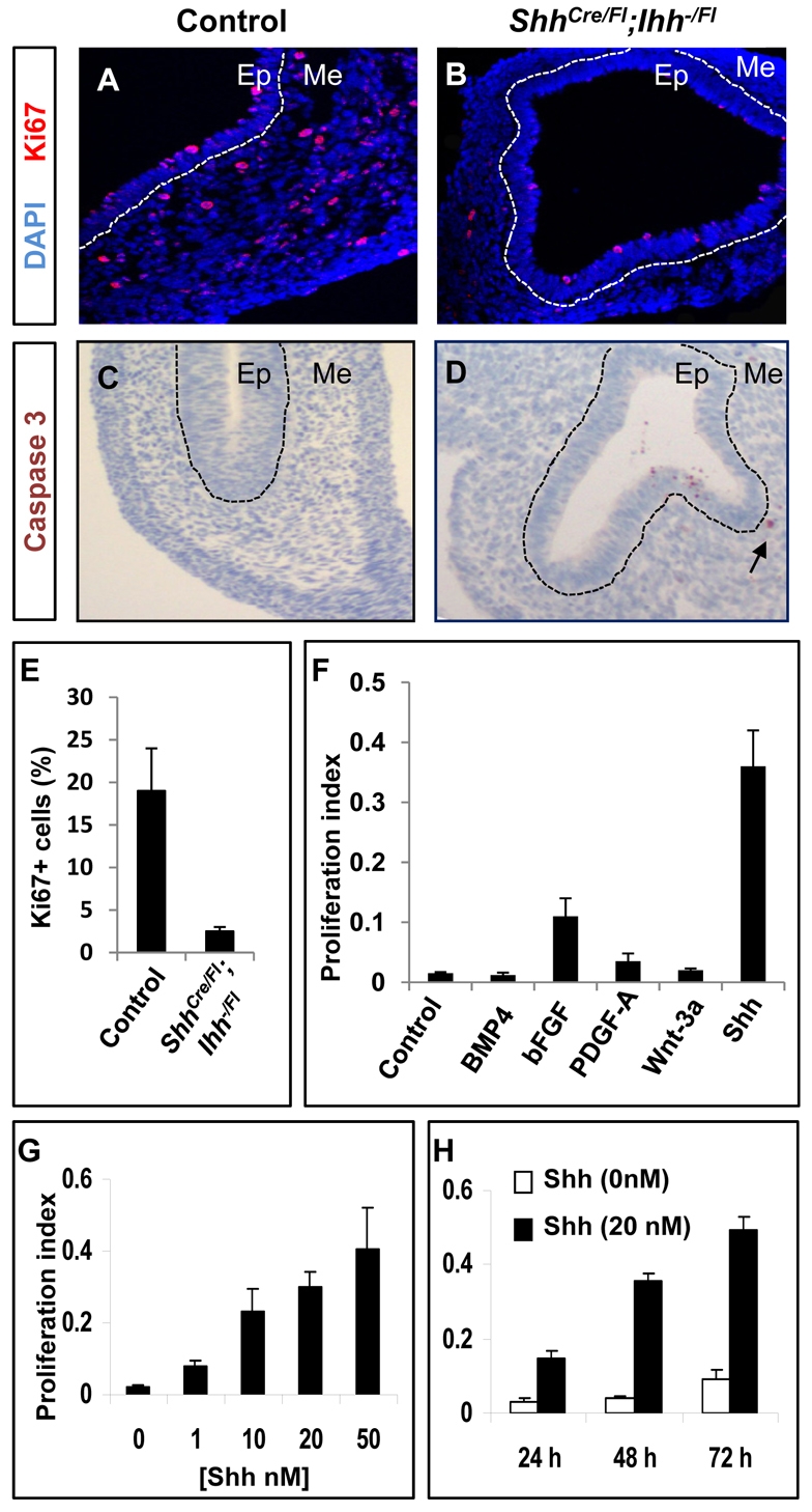
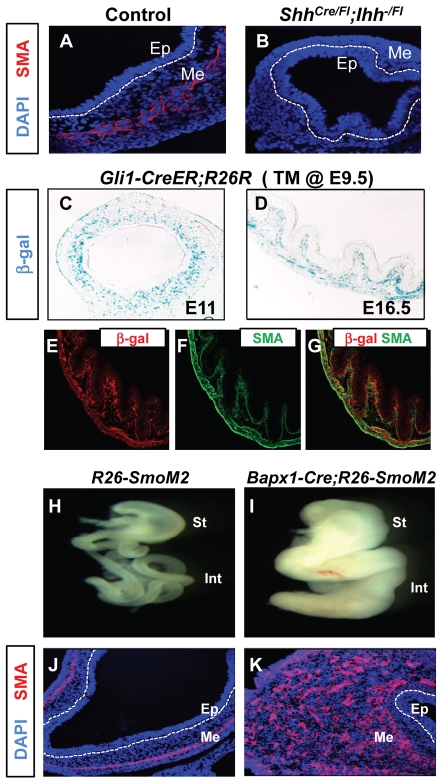
Homeostasis of the vertebrate digestive tract requires interactions between an endodermal epithelium and mesenchymal cells derived from the splanchnic mesoderm. Signaling between these two tissue layers is also crucial for patterning and growth of the developing gut. From early developmental stages, sonic hedgehog (Shh) and indian hedgehog (Ihh) are secreted by the endoderm of the mammalian gut, indicative of a developmental role. Further, misregulated hedgehog (Hh) signaling is implicated in both congenital defects and cancers arising from the gastrointestinal tract. In the mouse, only limited gastrointestinal anomalies arise following removal of either Shh or Ihh. However, given the considerable overlap in their endodermal expression domains, a functional redundancy between these signals might mask a more extensive role for Hh signaling in development of the mammalian gut. To address this possibility, we adopted a conditional approach to remove both Shh and Ihh functions from early mouse gut endoderm. Analysis of compound mutants indicates that continuous Hh signaling is dispensable for regional patterning of the gut tube, but is essential for growth of the underlying mesenchyme. Additional in vitro analysis, together with genetic gain-of-function studies, further demonstrate that Hh proteins act as paracrine mitogens to promote the expansion of adjacent mesenchymal progenitors, including those of the smooth muscle compartment. Together, these studies provide new insights into tissue interactions underlying mammalian gastrointestinal organogenesis and disease.


Figures

Fig. 1.
Fig. 1.
Removal of Shh and Ihh activities in the developing mouse gut. (A-G) Dynamic Shh and Ihh expression in the gut. Wholemount (A,B,D-F) and tissue-section (C,G) in situ hybridization for Shh (A-C) and Ihh (D-G) in wild-type mouse embryos from E10.5 to E12.5, showing coexpression of Hh ligands and partial overlap in the embryonic stomach (St) and intestine (Int), including small intestine (Sm Int) and large intestine (La Int). A, B, D and F show frontal views of the isolated digestive tract; lung buds (Lu) are included in A, D and E. Inset in E highlights the lack of Ihh expression in the lung buds, esophagus (Es) and rostral stomach. Distinctive fore-stomach and hind-stomach expression is shown in the photomicrographs in C and G. (H,I) Wholemount staining for β-gal in progeny of ShhCre/+ and ROSA26R mice, illustrating efficient recombination throughout the aerodigestive tract at E10.5 (H) and E11.5 (I). Trachea and lung buds are present at the top of both wholemount specimens and blue signal is visible throughout the stomach and intestine. (J-Q) Gross gastrointestinal phenotypes in control and ShhCre/Fl;Ihh−/Fl mutant embryos. Wholemount images of the isolated digestive tract, including stomach and intestine, at E11.5, E12.5, E14.5 and E16.5. Lung buds and esophagus are shown at E11.5 (J,K). As embryos age, the stomach lumen stretches markedly, most likely as a result of an absence of mesenchyme-derived muscular support. The spleen (arrows) is reduced in size at E16.5 (P,Q).
Fig. 2.
Fig. 2.
Digestive tract growth and differentiation in ShhCre/Fl;Ihh−/Fl mutant embryos. (A) Histology of the developing stomach (St) and adjoining spleno (Sp)-pancreatic (Pa) primordium in Shh;Ihh double mutant (right) and control (compound Shh+/Fl;Ihh+/Fl or Shh+/Fl;Ihh−/Fl heterozygote littermates, left) embryos from E11.5 to E16.5. (B) High-magnification micrographs from Shh;Ihh double-mutant (right) and control (left) embryos showing mesenchymal expansion and smooth muscle differentiation in the latter. Progressive attrition of the mesenchymal compartment in mutants is the probable basis for failure of organ growth and lack of epithelial differentiation. Dotted white lines separate gut endodermal epithelium (Ep) from the underlying mesenchyme (Me). Scale bars: 150 μm in A (E16.5, 500 μm); 75 μm in B (E16.5, 500 μm).
Fig. 3.
Fig. 3.
Epithelial differentiation of ShhCre/Fl;Ihh−/Fl mutant gut. (A) Wholemount of the ShhCre/Fl;Ihh−/Fl digestive tract at E18.5, showing inflation of the stomach as a result of wall thinning. The spleen is reduced in size but liver dimensions are unchanged. (B-D) Histology of ShhCre/Fl;Ihh−/Fl stomach at E18.5, emphasizing loss of wall thickness and regions of limited squamous and glandular differentiation, shown at higher resolution in C and D, respectively. (E-G) The rudimentary stomach mucosa shows isolated areas of foveolar cell differentiation at E18.5, indicated by staining with Periodic Acid Schiff (PAS; E) and Muc5AC (F), largely confined to regions where a few mesenchymal cells remain. H/K-ATPase (H+K+), a parietal cell marker, is expressed in most mucosal cells (G), even when underlying mesenchyme is absent at E18.5. (H-J) Primitive villous differentiation in Shh;Ihh double-mutant intestine at E18.5. Very few intestinal villi (arrows in H) form in the absence of Hh signaling, mainly near residual mesenchymal cells; most of the intestinal epithelium remains flat (arrowheads in H). These few villi carry goblet cells, which stain with Alcian Blue (Alc Bl; I), and express mRNA for the enterocyte marker Apo1a (J). Pa, pancreatic tissue. (K-M) Delineation of gut anteroposterior (A-P) pattern in E12.5 ShhCre/Fl;Ihh−/Fl mutant mouse embryos, as judged by expression of regional epithelial markers. (K) p63, a squamous cell product, reveals a sharp boundary between stomach squamous and glandular mucosae. (L,M) Staining for Pdx1, which marks the stomach (St) antrum, duodenum (Duo) and pancreas (Pa), and the intestine-specific marker Cdx2 in consecutive E12.5 tissue sections further demonstrates correct regional gene expression in absence of Hh signaling.
Fig. 4.
Fig. 4.
Mesenchymal differentiation of ShhCre/Fl;Ihh−/Fl mutant gut. (A,B) Neuron-specific β-tubulin, Tuj1, expression in control and ShhCre/Fl;Ihh−/Fl mutant stomachs at E12.5. (C,D) Endothelial marker CD31 immunostaining at E12.5. (E,F) Endothelial marker Fli1 immunostaining in control and ShhCre/Fl;Ihh−/Fl mutant stomachs at E12.5. Dashed white lines separate gut endodermal epithelium (Ep) from the underlying mesenchyme (Me). (G,H) Erythrocytes containing blood vessels are present in the mesenchyme of ShhCre/Fl;Ihh−/Fl mutant stomachs at E12.5 (G) and E16.5 (H).
Fig. 5.
Fig. 5.
Hh ligands promote proliferation of fetal GI mesenchymal cells. (A,B) Ki67 immunostaining in E12.5 ShhCre/Fl;Ihh−/Fl mutant stomach shows reduced proliferative activity in mesenchymal cells relative to controls. Dashed white lines delineate epithelium (Ep) and mesenchyme (Me). (C,D) cleaved caspase 3 immunostaining in control and Hh mutant stomachs at E12.5. Arrow in D shows the positive caspase 3 staining in Hh mutant mesenchyme. (E) Graph of the percentage of Ki67-expressing mesenchymal cells per 40× field in control and ShhCre/Fl;Ihh−/Fl mutant E12.5 stomachs. Two mice were analyzed per genotype counting a minimum of eight fields per mouse. (F) Proliferative effects of different signaling molecules on fetal stomach mesenchymal cells. Isolated E12.5 stomach mesenchymal cells were cultured for 72 hours as indicated in the presence of N-Shh or Wnt3a conditioned medium, or recombinant Bmp4 (100 ng/ml), bFGF (25 ng/ml) or Pdgf-AA (25 ng/ml). (G,H) Stomach mesenchymal cells were isolated from wild-type E12.5 embryos and cultured in the presence of increasing concentrations (G) of recombinant N-Shh for 3 days, or for different periods in 20 mM N-Shh (H). Proliferative activity was measured by the CellTiter 96 Non-Radioactive Cell Proliferation Assay kit (F-H).
Fig. 6.
Fig. 6.
Hh pathway activation and smooth muscle development in GI mesenchyme. (A,B) The absence of smooth muscle actin (SMA) expression in ShhCre/Fl;Ihh−/Fl mutant stomachs at E12.5. (C-G) Hh-responding early progenitor cells give rise to smooth muscle cells and myofibrolasts in gut mesenchyme. β-gal staining (C,D) of E11 and E16.5 Gli1-CreER;R26R gastric mesenchyme following Cre activation by tamoxifen (TM) at E9.5. β-gal and SMA immunostaining (E-G) of Gli1-CreER;R26R gastric mesenchyme at E16.5. (H,I) Wholemount images of E14.5 GI tracts, including stomach (St) and intestine (Int), from R26-SmoM2 and Bapx1-Cre;R26-SmoM2 embryos. (J,K) Smooth muscle actin (SMA) expression in R26-SmoM2 and Bapx1-Cre;R26-SmoM2 embryonic gut at E14.5. SMA immunostaining shows marked expansion of the mesenchymal compartment, including SMA-expressing smooth muscle cells in the Bapx1-Cre;R26-Smom2 stomach mesenchyme. Dashed white lines delineate epithelium (Ep) and mesenchyme (Me).

References

    1. Ahn S., Joyner A. L. (2004). Dynamic changes in the response of cells to positive hedgehog signaling during mouse limb patterning. Cell 118, 505-516 - PubMed
    1. Apelqvist A., Ahlgren U., Edlund H. (1997). Sonic hedgehog directs specialised mesoderm differentiation in the intestine and pancreas. Curr. Biol. 7, 801-804 - PubMed
    1. Arsic D., Qi B. Q., Beasley S. W. (2002). Hedgehog in the human: a possible explanation for the VATER association. J. Paediatr. Child Health 38, 117-121 - PubMed
    1. Aubin J., Dery U., Lemieux M., Chailler P., Jeannotte L. (2002). Stomach regional specification requires Hoxa5-driven mesenchymal-epithelial signaling. Development 129, 4075-4087 - PubMed
    1. Bhowmick N. A., Neilson E. G., Moses H. L. (2004). Stromal fibroblasts in cancer initiation and progression. Nature 432, 332-337 - PMC - PubMed

Publication types

MeSH terms

Substances