. 2010 Sep;38(16):5554-68.
doi: 10.1093/nar/gkq305. Epub 2010 Apr 29.

ERAL1 is associated with mitochondrial ribosome and elimination of ERAL1 leads to mitochondrial dysfunction and growth retardation

Affiliations

ERAL1 is associated with mitochondrial ribosome and elimination of ERAL1 leads to mitochondrial dysfunction and growth retardation

Takeshi Uchiumi et al. Nucleic Acids Res. 2010 Sep.

Abstract

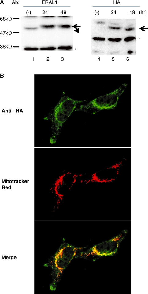
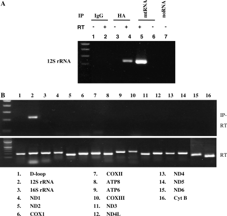
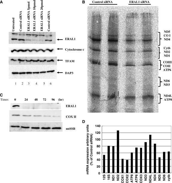
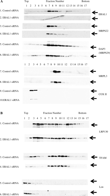
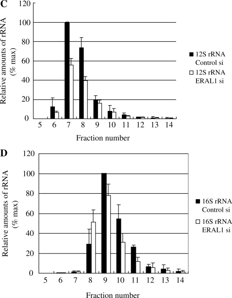
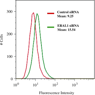
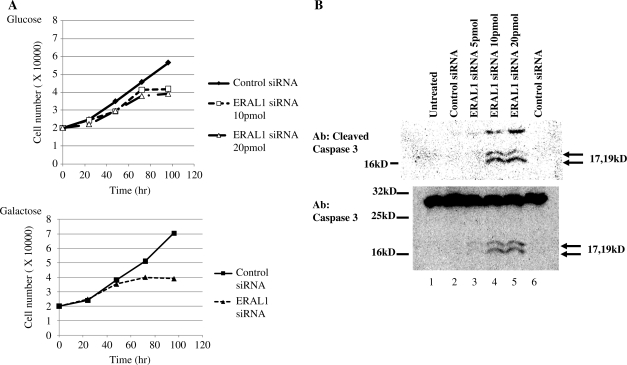
ERAL1, a homologue of Era protein in Escherichia coli, is a member of conserved GTP-binding proteins with RNA-binding activity. Depletion of prokaryotic Era inhibits cell division without affecting chromosome segregation. Previously, we isolated ERAL1 protein as one of proteins which were associated with mitochondrial transcription factor A by using immunoprecipitation. In this study, we analysed the localization and function of ERAL1 in mammalian cells. ERAL1 was localized in mitochondrial matrix and associated with mitoribosomal proteins including the 12S rRNA. siRNA knockdown of ERAL1 decreased mitochondrial translation, caused redistribution of ribosomal small subunits and reduced 12S rRNA. The knockdown of ERAL1 in human HeLa cells elevated mitochondrial superoxide production and slightly decreased mitochondrial membrane potential. The knockdown inhibited the growth of HeLa cells with an accumulation of apoptotic cells. These results suggest that ERAL1 is localized in a small subunit of the mitochondrial ribosome, plays an important role in the small ribosomal constitution, and is also involved in cell viability.


Figures

Figure 1.
Figure 1.
Inducible expression of recombinant ERAL1 with an HA tag. (A) ERAL1 with an HA tag was inducibly expressed in HeLa cells using a Tet-on system as described under ‘Methods’ section. ERAL1 was detected by immunoblotting with anti-ERAL1 (lanes 1–3) and anti-HA (lanes 4–6) antibodies. Lanes 1/4; no induction, lanes 2/5 and 3/6; 24 and 48 h after tetracycline treatment, respectively. Arrows and an arrowhead indicate recombinant and endogenous ERAL1, respectively. Asterisks show non-specific bands. (B) Immunocytochemistry of recombinant ERAL1. The ERAL1-HA-transfected cells were cultured in the presence of doxycyclin for 48 h. Mitochondria and the recombinant ERAL1 were visualized with a mitochondria-staining dye, MitoTracker Red, (middle panels) and anti-HA antibodies (upper panel), respectively. The lower panels are merged.
Figure 2.
Figure 2.
Submitochondrial localization of exogenous ERAL1-HA. Mitochondria were incubated in hypotonic buffer for disruption of the outer membranes without (lanes 3 and 4) or with a non-ionic detergent, Triton X-100 (lane 5). Then the mitochondria were digested with proteinase K (lanes 2, 4 and 5). The indicated proteins were detected by immunoblotting. EndoG, BAP37 and TFAM are markers for the mitochondrial IMS, IM and matrix, respectively.
Figure 3.
Figure 3.
ERAL1 is associated with mitochondrial 12S rRNA in HeLa cells. (A) ERAL1-HA-transfected HeLa cells were treated by tetracycline for 48 h. The cells were lysed by TNE buffer and immunoprecipitated with control IgG (lanes 1 and 2) or anti-HA antibodies (lanes 3 and 4). RNAs were extracted using an RNAeasy kit from the immunoprecipitates (lanes 1–4) and whole cells (lanes 5 and 6) and then treated with DNaseI. The DNaseI-treated RNAs were subjected to RT (lanes 2, 4 and 5) or not subjected to RT (lanes 1, 3 and 6) and then amplified by PCR with a primer set for 12S rRNA. No RNA template in lane 7. (B) The RNA in lane 4 of (A) (IP-RT, upper panel) and that in lane 5 of (A) (RT, lower panel) were amplified similarly by PCR using indicated primer sets for 16 mitochondrial genes.
Figure 4.
Figure 4.
Mitochondrial proteins crosslinked to ERAL1-HA. ERAL1-HA-transfected HeLa cells were treated by tetracycline for 48 h. Mitochondrial fraction was purified by percoll density gradient and lysed by TNE buffer. Immunoprecipitates obtained with anti-HA and mouse-IgG were separated by SDS–PAGE and then immunoblotted with anti-EF-Tu, ERAL1, LRP130, TFAM, mtSSB and DAP3 antibodies. An asterisk indicates a heavy chain of IgG.
Figure 5.
Figure 5.
siRNA-mediated knockdown of ERAL1 and in vivo labelling. (A) HeLa cells were transfected with nothing (lane 1), control siRNA (lanes 2 and 6), and 5, 10, 20 pmol of ERAL1 siRNA (lanes 3–5) using oligofectamine. After 2 days of the transfection, the cells were lysed and followed by immunoblotting with anti-ERAL1, cytochrome c, TFAM and DAP3 antibodies. (B) Synthesis of mitochondrial polypeptides. HeLa cells were transfected with control and ERAL1 siRNAs for 72 h. These cells were pulse-labelled in the presence of emetine. The mitochondrial translation products were run on a 15–20% polyacrylamide gradient gel. The gel was processed for fluorography as detailed in the ‘Methods’ section. The seven complex I subunits (ND), one complex III subunit (cyt b), three complex IV subunits (COX) and two complex V subunits (ATP) are indicated on the right. (C) siRNA-mediated knockdown of ERAL1. HeLa cells were transfected with ERAL1 siRNA and harvested every 24 h. The cells were lysed and followed by immunoblotting with anti-ERAL1, COX II and mtSSB antibodies. (D) Quantification of mitochondria-encoded mRNA. HeLa cells were treated with control or ERAL1 siRNA for 72 h. The amounts of mRNAs were measured as described under ‘Methods’ section. The value in control siRNA-treated cells is 100% for each mRNA.
Figure 6.
Figure 6.
Reduction of ERAL1 results in redistribution of ribosomal protein and ribosomal RNA. (A and B) Cell lysates were prepared from HeLa cells treated with ERAL1 or non-targeted (control) siRNA for 72 h. After separation by centrifugation through 10–35% sucrose, the sample was divided into 17 fractions. Each fraction was analysed by western blotting using antibodies against the small (DAP3, MRPS22) and large (MRPL3) mitochondrial ribosomal subunits, ERAL1, COX II, TFAM, Lon and LRP130. (C and D) Ribosomal RNA levels of each fraction were analyzed by using real time PCR: 12S mitochondrial ribosomal RNA (C), 16S mitochondrial ribosomal RNA (D). The maximal value is 100% for each RNA.
Figure 6.
Figure 6.
Reduction of ERAL1 results in redistribution of ribosomal protein and ribosomal RNA. (A and B) Cell lysates were prepared from HeLa cells treated with ERAL1 or non-targeted (control) siRNA for 72 h. After separation by centrifugation through 10–35% sucrose, the sample was divided into 17 fractions. Each fraction was analysed by western blotting using antibodies against the small (DAP3, MRPS22) and large (MRPL3) mitochondrial ribosomal subunits, ERAL1, COX II, TFAM, Lon and LRP130. (C and D) Ribosomal RNA levels of each fraction were analyzed by using real time PCR: 12S mitochondrial ribosomal RNA (C), 16S mitochondrial ribosomal RNA (D). The maximal value is 100% for each RNA.
Figure 7.
Figure 7.
Increased ROS production by ERAL1 siRNA-mediated knockdown. HeLa cells were transfected with control (Green) and ERAL1 siRNAs (Red). After 72 h, the cells were treated 10 µM H2DCFDA for 30 min and were subjected to FACS analysis for quantitative estimation of ROS.
Figure 8.
Figure 8.
Depletion of ERAL1 caused a growth defect and increased apoptosis. (A) Proliferation rate of siRNA-transfected HeLa cells. The cells were cultured with a normal glucose medium (upper) or a galactose medium (lower). HeLa cells were transfected with control and ERAL1 siRNAs at Day 0. At indicated time, the cells were harvested and counted by a cell counter. (B) HeLa cells were transfected with control and ERAL siRNAs for 72 h. Untransfected (lane1) and transfected HeLa cells were lysed and immunoblotted by anti-cleaved caspase 3 (upper panel) and anti-caspase 3 (lower panel) antibodies, respectively.

References

    1. Kang D, Hamasaki N. Mitochondrial transcription factor A in the maintenance of mitochondrial DNA: overview of its multiple roles. Ann. NY Acad. Sci. 2005;1042:101–108. - PubMed
    1. Asin-Cayuela J, Gustafsson CM. Mitochondrial transcription and its regulation in mammalian cells. Trends Biochem. Sci. 2007;32:111–117. - PubMed
    1. Falkenberg M, Larsson NG, Gustafsson CM. DNA replication and transcription in mammalian mitochondria. Annu. Rev. Biochem. 2007;76:679–699. - PubMed
    1. Pel HJ, Grivell LA. Protein synthesis in mitochondria. Mol. Biol. Rep. 1994;19:183–194. - PubMed
    1. Rorbach J, Soleimanpour-Lichaei R, Lightowlers RN, Chrzanowska-Lightowlers ZM. How do mammalian mitochondria synthesize proteins? Biochem. Soc. Trans. 2007;35:1290–1291. - PubMed

Publication types

MeSH terms