. 2010 Jun 25;285(26):19927-34.
doi: 10.1074/jbc.M110.126581. Epub 2010 Apr 29.

The acetyltransferase activity of the bacterial toxin YopJ of Yersinia is activated by eukaryotic host cell inositol hexakisphosphate

Affiliations

The acetyltransferase activity of the bacterial toxin YopJ of Yersinia is activated by eukaryotic host cell inositol hexakisphosphate

Rohit Mittal et al. J Biol Chem. .

Abstract

Plague, one of the most devastating diseases in human history, is caused by the bacterium Yersinia pestis. The bacteria use a syringe-like macromolecular assembly to secrete various toxins directly into the host cells they infect. One such Yersinia outer protein, YopJ, performs the task of dampening innate immune responses in the host by simultaneously inhibiting the MAPK and NFkappaB signaling pathways. YopJ catalyzes the transfer of acetyl groups to serine, threonine, and lysine residues on target proteins. Acetylation of serine and threonine residues prevents them from being phosphorylated thereby preventing the activation of signaling molecules on which they are located. In this study, we describe the requirement of a host-cell factor for full activation of the acetyltransferase activity of YopJ and identify this activating factor to be inositol hexakisphosphate (IP(6)). We extend the applicability of our results to show that IP(6) also stimulates the acetyltransferase activity of AvrA, the YopJ homologue from Salmonella typhimurium. Furthermore, an IP(6)-induced conformational change in AvrA suggests that IP(6) acts as an allosteric activator of enzyme activity. Our results suggest that YopJ-family enzymes are quiescent in the bacterium where they are synthesized, because bacteria lack IP(6); once injected into mammalian cells by the pathogen these toxins bind host cell IP(6), are activated, and deregulate the MAPK and NFkappaB signaling pathways thereby subverting innate immunity.


Figures

FIGURE 1.
FIGURE 1.
Assays for the acetylation of MEK; eukaryotic HeLa cells contain an activating cofactor for YopJ. A, HeLa cells were transfected with wild-type YopJ (pSFFV-YopJ wt) or with the enzymatically inactive C172A mutant (pSFFV-YopJ C172A). Cell lysates were immunoblotted for endogenous MEK using an antiserum that is sensitive to acetylation of MEK (CST9122) and another that is not sensitive to the modification (47E6). It is seen that expression of wild-type YopJ leads to modification of the majority of endogenous MEK. B, an in vitro acetylation assay carried out in the presence of [14C]AcCoA shows that progressively higher amounts of YopJ (0.3–1.2 μg) led to an increase in the acetylation of MEK (5 μg). C, inclusion of HeLa cell cytosol (1 μl of a 3 mg/ml preparation) results in the stimulation of YopJ acetyltransferase activity as evidenced by the increased acetylation of MEK and also the increased autoacetylation of YopJ (in lane 3 compared with lane 2).
FIGURE 2.
FIGURE 2.
Analysis of the cofactor activity from HeLa cytosol; absence of cofactor activity in bacteria. A, Fraction 52 obtained from size-exclusion chromatography of HeLa cytosol (supplemental Fig. S1) was used in the in vitro acetyltransferase assay and displayed significant stimulation of YopJ activity (compare lane 3 to lane 2). Heating Fraction 52 at 95 °C for 30 min did not destroy the cofactor activity (compare lane 4 to lane 3). B, the activating cofactor for YopJ was also present in an acid extract prepared from HeLa cells (lane 4). C, acid extracts (AE) prepared from the bacteria E. coli, yeast S. cerevisiae, amoeba (D. discoideum), and mammalian sources, HeLa cells and rat brain lysate (RBL), were tested for the presence of cofactor activity by assessing their ability to stimulate the acetylation of MEK by YopJ in vitro. Of the extracts tested, only the bacterial extract (lane 4) was devoid of cofactor activity whereas the extracts prepared from different eukaryotic sources displayed presence of the activity (lanes 5–8). Fraction 52 (lane 3) was used as the positive control.
FIGURE 3.
FIGURE 3.
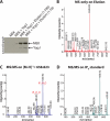
Cofactor activity could be purified by anion exchange chromatography and was identified by MS to be IP6. A, cofactor activity present in the HeLa cell acid extract was eluted from AG1-X8 resin, lyophilized, and resuspended in water for inclusion in acetyltransferase assays. The stimulation of YopJ activity by two different dilutions (1:100 and 1:10) of the elution is shown here. The elution was then subjected to mass spectrometric analysis. B, MALDI-MS spectrum acquired in the reflectron negative ion mode revealed a prominent m/z peak of 658.823 Da. This peak was manually selected for further MS/MS fragmentation (C), and the resulting ion series corresponds to the breakdown products expected for IP6. D, MS/MS fragmentation ion series obtained for an IP6 standard.
FIGURE 4.
FIGURE 4.
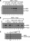
Validation of IP6 as the activating cofactor. A, autoradiograph examining acetylation (using [14C]AcCoA) of MEK by YopJ in the presence of increasing doses (1, 10, and 100 nm) of IP3, IP6, and IS6. In A and B, lane 1 depicts the acetylation of MEK by YopJ in the absence of any added stimulatory factor. It is seen in panel A that only IP6 (lanes 5–7) causes a stimulation of YopJ activity. B, the stimulation caused by IP6 (lane 2) is reversed by the addition of phytase (Phy, lane 3) but not by alkaline phosphatase (AP, lane 4). Phytase sensitivity is also seen for the stimulatory activity present in the HeLa cell acid extract (HAE) shown here for two different dilutions (1:100 and 1:10) of the extract. C, a YopJ-catalyzed acetyltransferase assay was performed (using non-radioactive AcCoA) with MEK as substrate in the absence or presence of IP6 or in the presence of increasing doses of HeLa acid extract (HAE). The reactions were then visualized by Western blot using the modification-sensitive anti-MEK antiserum (CST9122). It is seen that acetylation of MEK by YopJ in the presence of IP6 results in almost complete loss of detection by CST9122 (compare lanes 1 and 2). Also, inclusion of progressively increasing amounts of the HAE in the acetylation reaction result in successively greater modification of MEK as evidenced by the loss of CST9122 immunodetection (lanes 3–7).
FIGURE 5.
FIGURE 5.
An inositol phosphate-deficient deletion strain of yeast lacks the cofactor; AvrA is stimulated by IP6 undergoing conformational change in response to IP6 addition. A, neutralized acid extracts were prepared from various deletion strains of the yeast S. cerevisiae and included in an acetyltransferase assay using [14C]AcCoA. Lane 1 shows the basal level of MEK acetylation catalyzed by YopJ. Inclusion of yeast extracts results in stimulation of the acetyltransferase activity of YopJ. Extract from the yeast strain ipmkΔ, deleted for the enzyme inositol phosphate multikinase that accumulates IP2 and IP3, does not have the ability to stimulate YopJ (lane 6). The predominant inositol polyphosphate species present in the extracts of the yeast strains used are also indicated in panel A. The stimulation resulting from the inclusion of 100 nm IP6 is shown as a positive control (in lane 8). As in earlier panels, note the concurrent increase in autoacetylation upon stimulation of YopJ. B, inclusion of IP6 (0–100 nm) results in increased autoacetylation of AvrA (1 μg), the YopJ homologue from S. typhimurium. C, change in the emission of protein tryptophan fluorescence of AvrA upon the addition of IP6 (final concentration, 10 μm) to AvrA (5.6 μm). D, change in ellipticity at 222 nm in the far UV CD spectrum of AvrA (5 μm) upon addition of IP6 (10 μm).

Similar articles

Cited by

References

    1. Mueller C. A., Broz P., Cornelis G. R. (2008) Mol. Microbiol. 68, 1085–1095 - PubMed
    1. Trosky J. E., Liverman A. D., Orth K. (2008) Cell Microbiol. 10, 557–565 - PubMed
    1. Monack D. M., Mecsas J., Ghori N., Falkow S. (1997) Proc. Natl. Acad. Sci. U.S.A. 94, 10385–10390 - PMC - PubMed
    1. Palmer L. E., Hobbie S., Galán J. E., Bliska J. B. (1998) Mol. Microbiol. 27, 953–965 - PubMed
    1. Mukherjee S., Keitany G., Li Y., Wang Y., Ball H. L., Goldsmith E. J., Orth K. (2006) Science 312, 1211–1214 - PubMed

Publication types

MeSH terms