. 2010 Jul 16;285(29):22114-21.
doi: 10.1074/jbc.M110.110361. Epub 2010 Apr 29.

CLOCK regulates circadian rhythms of hepatic glycogen synthesis through transcriptional activation of Gys2

Affiliations

CLOCK regulates circadian rhythms of hepatic glycogen synthesis through transcriptional activation of Gys2

Ryosuke Doi et al. J Biol Chem. .

Abstract

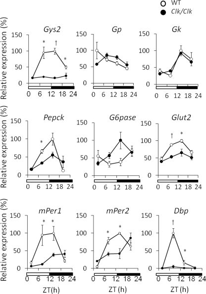
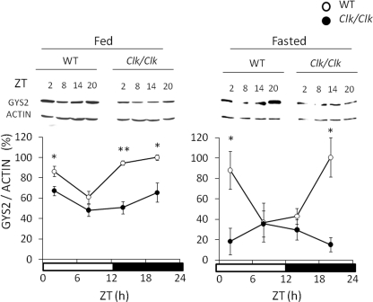
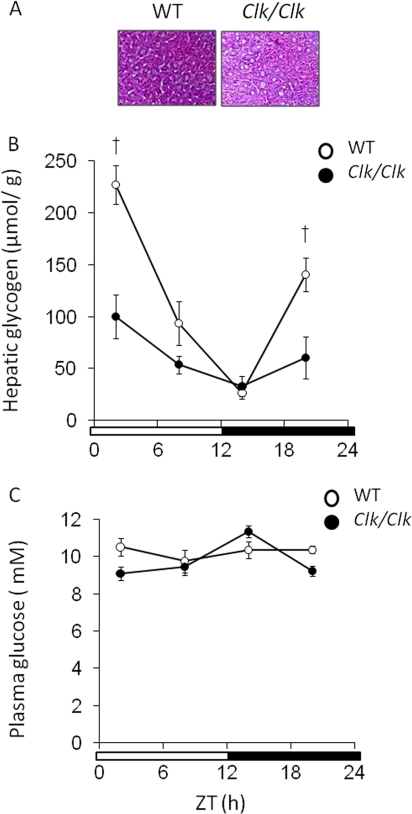
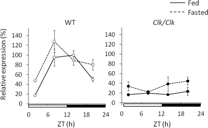
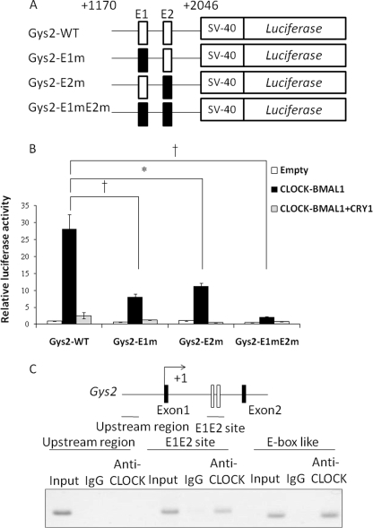
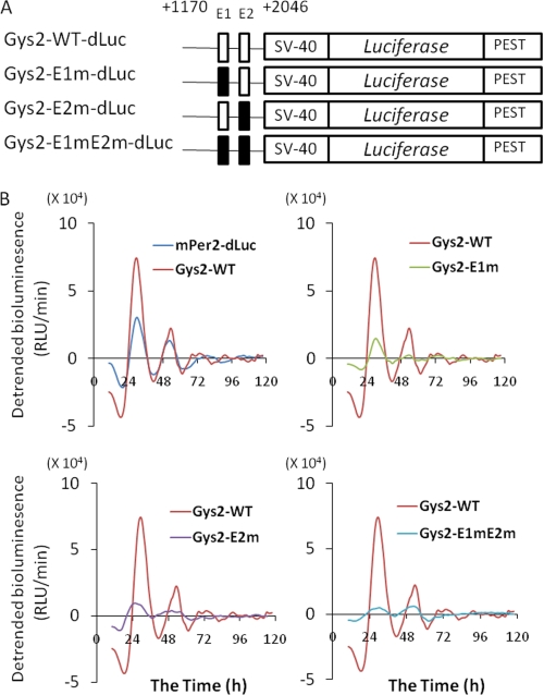
Hepatic glycogen content is important for glucose homeostasis and exhibits robust circadian rhythms that peak at the end of the active phase in mammals. The activities of the rate-limiting enzymes for glycogenesis and glycogenolysis also show circadian rhythms, and the balance between them forms the circadian rhythm of the hepatic glycogen content. However, no direct evidence has yet implicated the circadian clock in the regulation of glycogen metabolism at the molecular level. We show here that a Clock gene mutation damps the circadian rhythm of the hepatic glycogen content, as well as the circadian mRNA and protein expression of Gys2 (glycogen synthase 2), which is the rate-limiting enzyme of glycogenesis in the liver. Transient reporter assays revealed that CLOCK drives the transcriptional activation of Gys2 via two tandemly located E-boxes. Chromatin immunoprecipitation assays of liver tissues revealed that CLOCK binds to these E-box elements in vivo, and real time reporter assays showed that these elements are sufficient for circadian Gys2 expression in vitro. Thus, CLOCK regulates the circadian rhythms of hepatic glycogen synthesis through transcriptional activation of Gys2.


Figures

FIGURE 1.
FIGURE 1.
Clock mutation affects the temporal expression of genes associated with glucose metabolism. The mice were maintained under a 12 h of light/12 h of dark cycle (lights on at 0:00 and lights off at 12:00). Total RNA was extracted from the livers of wild-type (WT) and Clock mutant mice, and the mRNA levels were quantified by real time reverse transcription-PCR. Open and closed symbols, wild-type and Clock mutant mice, respectively. Amount of mRNA was corrected relative to that of β-actin. The maximal value for wild-type mice is expressed as 100%. The values are the means ± S.E. (n = 3). The significant differences compared with values from wild-type mice at each time point are indicated. *, p < 0.05; †, p < 0.01. The p values were calculated using Student's t test.
FIGURE 2.
FIGURE 2.
Clock mutation damps circadian variation of GYS2 protein in both fed and fasted mice. Wild-type (WT) and Clock mutant mice were maintained under a 12 h of light/12 h of dark cycle (lights on at 0:00 and lights off at 12:00) and fed (left panel) or fasted (right panel) overnight. Protein was extracted from the livers of these mice, and then the GYS2 protein level was determined by Western blotting using anti-GYS2 antiserum. Open and closed symbols, wild-type and Clock mutant mice, respectively. The maximal value for wild-type mice is expressed as 100%. The values are the means ± S.E. (n = 3). The significant differences compared with the values from wild-type mice at each time point are indicated. *, p < 0.05; †, p < 0.01. The p values were calculated using Student's t test.
FIGURE 3.
FIGURE 3.
Clock mutation damps circadian variation of hepatic glycogen contents. The mice were maintained under a 12 h of light/12 h of dark cycle (lights on at 0:00 and lights off at 12:00). Open and closed symbols, wild-type (WT) and Clock mutant mice, respectively. A, periodic acid-Schiff's staining of liver sampled at ZT 2 of wild-type mice and Clock mutant mice (n = 3). B, hepatic glycogen contents in wild-type mice and Clock mutant mice were measured at indicated times. The values are the means ± S.E. (n = 8). C, plasma glucose concentrations. The values are the means ± S.E. (n = 8). The significant differences compared with values from wild-type mice at each time point are indicated. †, p < 0.01. The p values were calculated using Student's t test.
FIGURE 4.
FIGURE 4.
Circadian expression of Gys2 continues during fasting. Wild-type (WT) and Clock mutant mice were maintained under a 12 h of light/12 h of dark cycle (lights on at 0:00 and lights off at 12:00) and fed (left panel) and fasted (right panel) overnight. Total RNA extracted from the livers of these mice was simultaneously quantified by real time reverse transcription-PCR. The data from fed mice of both genotypes are identical to those shown in Fig. 1. Open and closed symbols, wild-type and Clock mutant mice, respectively. The solid and dotted lines indicate fed and fasted mice, respectively. The amount of mRNA was corrected relative to that of β-actin. The maximal value for wild-type mice is expressed as 100%. The values are the means ± S.E. (n = 3).
FIGURE 5.
FIGURE 5.
CLOCK drives transcriptional activation of Gys2 via two E-boxes in the first intron. A, schematic representation of mutant constructs of Gys2 first intronic region. Each or both E-boxes (sites E1 and E2) were mutated (E1 site, 5′-CACGTG-3′ to 5′-CACCAC-3′; E2 site, 5′-CACGTG-3′ to 5′-CACCAC-3′). B, analysis of E1 and E2 sites in the first Gys2 intronic region. Transcriptional assays included the indicated mutant constructs. Gys2-WT, wild type of first intron on Gys2 gene; Gys2-E1m, mutant E1 site; Gys2-E2m, mutant E2 site; Gys2-E1mE2m, mutant E1 and E2 sites. The normalized expression level was calculated relative to luciferase activity in empty vector. The values are described as the means ± S.E. (n = 3). Significant differences are indicated. *, p < 0.05; †, p < 0.01. p values were calculated using Student's t tests. The results are representative of three independent experiments. C, CLOCK binds to the first intronic region containing E1 and E2 sites in vivo. The chromatin immunoprecipitation assays were performed using livers from mice at ZT14. The region containing two E-boxes is shown as an open box. Horizontal bars, amplified regions (upstream region, from −2864 to −2620; E1E2 site on first intron, from +1541 to +1804). The E-box (CACGTT) on Per2 promoter known as CLOCK binding region served as a positive control. IgG, with mouse normal IgG; Anti-CLOCK, with anti-CLOCK antibody.
FIGURE 6.
FIGURE 6.
Sites E1 and E2 are sufficient for circadian expression of Gys2. A, schematic representation of mutant constructs of Gys2 first intronic region. Each or both E-boxes (sites E1 and E2) were mutated (E1, 5′-CACGTG-3′ to 5′-CACCAC-3′; E2, 5′-CACGTG-3′ to 5′-CACCAC-3′). B, real time reporter assay. NIH3T3 cells were transfected with indicated mutant constructs and incubated with 100 nm of dexamethasone, and then bioluminescence was measured. The results are representative of three independent experiments that generated similar outcomes. Gys2-WT-dLuc, wild type of first intron on Gys2 gene; Gys2-E1m-dLuc, mutant E1 site; Gys2-E2m-dLuc, mutant E2 site; Gys2-E1mE2m-dLuc, mutant E1 and E2 sites (see “Experimental Procedures” for details of methods for analyzing circadian rhythms).

References

    1. Pando M. P., Sassone-Corsi P. (2001) Nature 410, 311–313 - PubMed
    1. Reppert S. M., Weaver D. R. (2001) Annu. Rev. Physiol. 63, 647–676 - PubMed
    1. Vitaterna M. H., King D. P., Chang A. M., Kornhauser J. M., Lowrey P. L., McDonald J. D., Dove W. F., Pinto L. H., Turek F. W., Takahashi J. S. (1994) Science 264, 719–725 - PMC - PubMed
    1. King D. P., Zhao Y., Sangoram A. M., Wilsbacher L. D., Tanaka M., Antoch M. P., Steeves T. D., Vitaterna M. H., Kornhauser J. M., Lowrey P. L., Turek F. W., Takahashi J. S. (1997) Cell 89, 641–653 - PMC - PubMed
    1. Sakamoto K., Nagase T., Fukui H., Horikawa K., Okada T., Tanaka H., Sato K., Miyake Y., Ohara O., Kako K., Ishida N. (1998) J. Biol. Chem. 273, 27039–27042 - PubMed

Publication types