. 2010 Jul;109(1):171-88.
doi: 10.1152/japplphysiol.00934.2009. Epub 2010 Apr 29.

Site-specific effects on respiratory rhythm and pattern of ibotenic acid injections in the pontine respiratory group of goats

Affiliations

Site-specific effects on respiratory rhythm and pattern of ibotenic acid injections in the pontine respiratory group of goats

J M Bonis et al. J Appl Physiol (1985). 2010 Jul.

Abstract

To probe further the contributions of the rostral pons to eupneic respiratory rhythm and pattern, we tested the hypothesis that ibotenic acid (IA) injections in the pontine respiratory group (PRG) would disrupt eupneic respiratory rhythm and pattern in a site- and state-specific manner. In 15 goats, cannulas were bilaterally implanted into the rostral pontine tegmental nuclei (RPTN; n = 3), the lateral (LPBN; n = 4) or medial parabrachial nuclei (MPBN; n = 4), or the Kölliker-Fuse nucleus (KFN; n = 4). After recovery from surgery, 1- and 10-microl injections (1 wk apart) of IA were made bilaterally through the implanted cannulas during the day. Over the first 5 h after the injections, there were site-specific ventilatory effects, with increased (P < 0.05) breathing frequency in RPTN-injected goats, increased (P < 0.05) pulmonary ventilation (Vi) in LPBN-injected goats, no effect (P < 0.05) in MPBN-injected goats, and a biphasic Vi response (P < 0.05) in KFN-injected goats. This biphasic response consisted of a hyperpnea for 30 min, followed by a prolonged hypopnea and hypoventilation with marked apneas, apneusis-like breathing patterns, and/or shifts in the temporal relationships between inspiratory flow and diaphragm activity. In the awake state, 10-15 h after the 1-microl injections, the number of apneas was greater (P < 0.05) than during other studies at night. However, there were no incidences of terminal apneas. Breathing rhythm and pattern were normal 22 h after the injections. Subsequent histological analysis revealed that for goats with cannulas implanted into the KFN, there were nearly 50% fewer neurons (P < 0.05) in all three PRG subnuclei than in control goats. We conclude that in awake goats, 1) IA injections into the PRG have site-specific effects on breathing, and 2) the KFN contributes to eupneic respiratory pattern generation.


Figures

Fig. 1.
Fig. 1.
Experimental design of the present study. Each goat underwent 2 surgeries, and on full recovery and training of goats, studies were conducted to establish the normal eupneic ventilatory parameters. These studies were repeated as shown to ascertain whether any perturbation affected the ventilatory parameters. MD, microdialysis; IA inj, ibotenic acid injection.
Fig. 2.
Fig. 2.
Histochemical and immunohistochemical staining for Nissl substance and muscarinic type-2 (M2) receptors provided anatomic characterization of the pontine respiratory group (PRG) in goats. A: sagittal sections 4.9 mm lateral to the midline showing the location in the rostral dorsal pons of the lateral (LPBN) and medial parabrachial nuclei (MPBN) and the Kölliker-Fuse nucleus (KFN) relative to the superior cerebellar peduncle (SCP). B: transverse Nissl and M2 sections and a schematic to further illustrate the location of the LPBN, MPBN, and KFN. Locations of the superior (SC) and inferior colliculus (IC), trigeminal motor nucleus (5), and basal pons (BP), facial nucleus (7), and nucleus ambiguus (NA) are indicated. D, dorsal; V, ventral; R, rostral; C, caudal; M, medial; L, lateral.
Fig. 3.
Fig. 3.
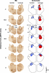
Immunohistochemical staining for M2 receptors of hemisections from 1 control goat and 1 goat that had a cannula implanted into the KFN. These hemisections illustrate the rostral-caudal changes in the LPBN, MPBN, and KFN beginning 1 mm caudal to the peak in number of KFN neurons (pKFN) and extending 3 mm rostral to the peak. The blue area in the schematic illustrates the orientation of the LPBN, MPBN, and KFN relative to the SCP. The tract of the cannula (gray area in schematic) extends over 2 mm, and the area devoid of neurons (red area in schematic) extends over 3 mm in the rostral-caudal direction. Note the clear presence of the KFN at its peak number of neurons (0 mm) and its absence at a more rostral distance (3 mm).
Fig. 4.
Fig. 4.
Number of neurons and intact area of control and lesioned (implanted) goats in the LPBN, MPBN, and KFN caudal and rostral to the pKFN. The red horizontal bars indicates values where neuron counts and the area of intact nuclei were reduced significantly (*P < 0.05) in the cannula-implanted goats. In the lesioned groups, the distal edge of the cannula was just dorsal to the LPBN, MPBN, or KFN. Note the 6-mm rostral-caudal extent of the LPBN and MPBN, whereas the KFN only extends over 3 mm.
Fig. 5.
Fig. 5.
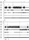
Periods of pursed-lipped breathing were associated with altered airway and pump muscle activity (B) compared with normal breathing (A). Note that in purse-lipped compared with normal breathing, there was high phasic abdominal (ABD) and thyroid arytenoid (TYA) muscle activity in late expiration, as well as high diaphragm (DIA) and genioglossus (GG) muscle activity, which appeared insufficient to maintain normal inspiratory flow (V̇i).
Fig. 6.
Fig. 6.
Both 1- and 10-μl bilateral injections of IA into the KFN resulted in arterial hypercapnia (PaCO2; hypoventilation) (*P < 0.05). Values shown are peak hypercapnia during the 5 h after the injections. IA injections into the RPTN, LPBN, and MPBN did not significantly (P > 0.05) affect PaCO2. In addition, PaCO2 was unchanged from the preimplant control at other times over the course of the experimental design.
Fig. 7.
Fig. 7.
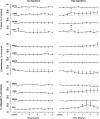
Bilateral injections of IA into the rostral pons of awake goats had heterogeneous, site-specific effects; bilateral injection of 1 or 10 μl of IA into the KFN had a biphasic response. First, pulmonary ventilation (V̇i) increased transiently (arrows), and second, V̇i was attenuated over several hours compared with this initial increase (†P < 0.05 compared with value at arrow). V̇i decreased due to a 37% decrease (*P < 0.05) in breathing frequency (f), despite an increase in tidal volume (Vt) (*P < 0.05). Bilateral injection of 10 μl of IA into the LPBN induced a persistent increase (*P < 0.05) in V̇i due to an increase in Vt, whereas injections into RPTN induced a delayed increase (*P < 0.05) in f. Bilateral injection of IA into the MPBN had no significant effect on breathing.
Fig. 8.
Fig. 8.
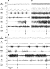
In a KFN-lesioned goat, bilateral injection of IA (10 μl) during the day caused a biphasic response in V̇i, blood pressure (BP), and heart rate compared with control conditions (A), with a transient (<20 min) hyperpnea (B) immediately after the injection, followed by a prolonged (>3 h) hypopnea (C). Arrows denote typical swallow patterns as identified by the abrupt, maximal contraction of the airway thyropharyngeus (TP) and TYA muscles. Electromyograms (EMG) are shown for the DIA, ABD, GG, TP, and TYA muscles. The post mortem histology of this goat's PRG is illustrated in Fig. 3.
Fig. 9.
Fig. 9.
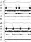
In a second KFN-lesioned goat, bilateral injection of IA (10 μl) during the day also caused a biphasic response in V̇i, BP, and heart rate compared with control conditions (A), with a transient (<20 min) hyperpnea (B) immediately after the injection, followed by a prolonged (>4 h) hypopnea (C). Arrows denote typical swallow patterns. Note in C the decrementing airflow pattern, decrementing GG muscle activity pattern, and increased ABD and TP activity that begin immediately after inspiration. PCA, posterior cricoarytenoid muscle activity.
Fig. 10.
Fig. 10.
In the second KFN-lesioned goat, prolonged expiratory time (central apnea) and/or a paired breathing pattern (A and B) occurred during the secondary hypopneic response to bilateral injection (10 μl) of IA. Note the decrementing airflow pattern, decrementing GG muscle activity, high level of ABD activity, and the change in BP during each inspiration except the second of a doublet inspiration.
Fig. 11.
Fig. 11.
In the second KFN-lesioned goat, apneusis-like breathing patterns (A and B) occurred during the secondary hypopneic response to bilateral injection (10 μl) of IA. During apneusis-like patterns (record between vertical lines), note the sustained DIA activity, which was terminated during phasic contraction of the ABD muscle. An upward arrow denotes a typical swallow pattern.
Fig. 12.
Fig. 12.
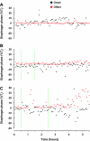
In the second KFN-lesioned goat, bilateral injection (ipsi- and contralateral injections made separately) of 10 μl of IA (vertical green lines) increased postinspiratory activity of the diaphragm, particularly 4–5 h after the 10-μl injections (C). Data in A and B were obtained during the control study and after injection of 1 μl of IA, respectively. Black symbols represent the delay in onset of airflow after the initiation of diaphragm activity, and the red symbols represent the delay in offset of diaphragm activity after cessation of inspiratory airflow. The delays in onset and offset are expressed as a percentage of inspiratory airflow time (Ti).
Fig. 13.
Fig. 13.
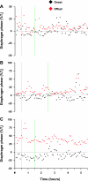
Similar to data shown in Fig. 12, in the other 3 KFN-lesioned goats (A–C), bilateral injections (ipsi- and contralateral injections made separately) of IA (vertical green lines) increased postinspiratory activity of the diaphragm during different periods after the injections. See Fig. 12 legend for further explanations.
Fig. 14.
Fig. 14.
Heart rate (HR) and mean arterial blood pressure (MABP) were significantly (*P < 0.05) increased and decreased, respectively, during portions of the 5-h period after bilateral 10-μl injections of IA into the KFN. Note the nonsignificant decrease in HR after the 1-μl injection. Neither HR nor MABP changed after injections into other rostral pontine areas.
Fig. 15.
Fig. 15.
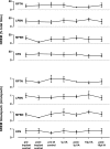
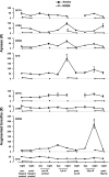
Over the course of the experimental protocol, the percentage of the night spent in non-rapid eye movement (NREM) sleep and the duration of sleep epochs did not change significantly in the RPTN, LPBN, MPBN, or KFN.
Fig. 16.
Fig. 16.
On the night of 1-μl injections of IA, the number of apneas was increased during wakefulness in KFN-lesioned goats (P < 0.05) and during NREM sleep in LPBN-lesioned goats (P < 0.001). Note that the number of apneas was nonsignificantly elevated in LPBN- and MPBN-lesioned goats during wakefulness but was not elevated whatsoever in animals with IA injections into RPTN. On the night of 10-μl injections of IA, the number of augmented breaths was increased during wakefulness in MPBN animals (P < 0.001). Asterisks denote significant differences from preimplant control values.
Fig. 17.
Fig. 17.
In a KFN-lesioned goat, unilateral injections of N-methyl-d-aspartate (NMDA) were made ipsi- and contralaterally the week before the first IA injection and the week after the last IA injection. The arrows indicate the time of the NMDA injection (1 μl, 100 mM). Compared with pre-IA NMDA injections, the magnitude of the hyperpnea was relatively preserved in post-IA NMDA injections, whereas the time course was somewhat delayed. V̇i is expressed in 1-min bins.

Similar articles

Cited by

References

    1. Abdala A, Rybak I, Smith J, Zoccal D, Machado B, St. John W, Paton J. Multiple pontomedullary mechanisms of respiratory rhythmogenesis. Respir Physiol Neurobiol 168: 19– 25, 2009 - PMC - PubMed
    1. Abdala A, Rybak I, Smith J, Paton J. Adominal expiratory activity in the rat brainstem-spinal cord in situ: patterns, origins and implications for respiratory rhythm generation. J Physiol 587: 3539– 3559, 2009 - PMC - PubMed
    1. Bellingham M, Funk G. Cholinergic modulation of respiratory brain-stem neurons and its function in sleep-wake state determination. Clin Exp Pharmacol Physiol 27: 132– 137, 2000 - PubMed
    1. Bolser D, Poliacek I, Jakus J, Fuller D, Davenport P. Neurogenesis of cough, other airway defensive behaviors and breathing: a holarchical system? Respir Physiol Neurobiol 152: 255– 265, 2006 - PMC - PubMed
    1. Chamberlin N, Saper C. Topographic organization of respiratory responses to glutamate microstimulation of the parabrachial nucleus in the rat. J Neurosci 14: 6500– 6510, 1994 - PMC - PubMed

Publication types

Substances

LinkOut - more resources