

Bacterial lipoproteins can disseminate from the periphery to inflame the brain
- PMID: 20431027
- PMCID: PMC2877846
- DOI: 10.2353/ajpath.2010.091235
Bacterial lipoproteins can disseminate from the periphery to inflame the brain
Abstract
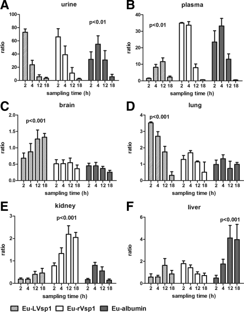
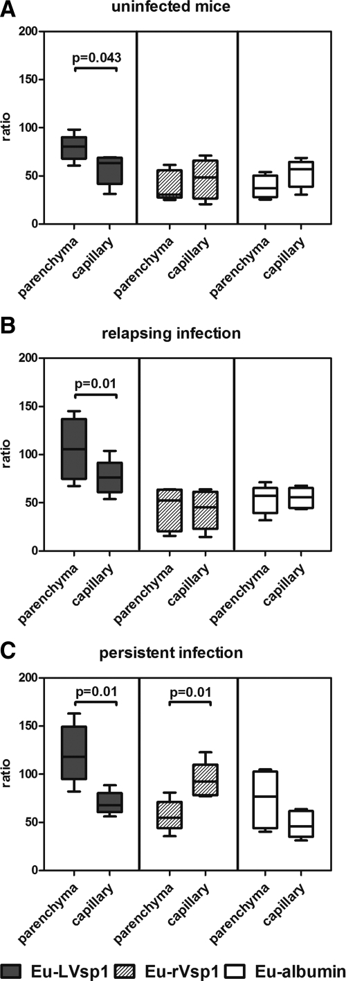
The current view is that bacteria need to enter the brain to cause inflammation. However, in mice infected with the spirochete Borrelia turicatae, we observed widespread cerebral inflammation despite a paucity of spirochetes in the brain parenchyma at times of high bacteremia. Here we studied the possibility that bacterial lipoproteins may be capable of disseminating from the periphery across the blood-brain barrier to inflame the brain. For this we injected normal and infected mice intraperitoneally with lanthanide-labeled variable outer membrane lipoproteins of B. turicatae and measured their localization in blood, various peripheral organs, and whole and capillary-depleted brain protein extracts at various times. Lanthanide-labeled nonlipidated lipoproteins of B. turicatae and mouse albumin were used as controls. Brain inflammation was measured by TaqMan RT-PCR amplification of genes known to be up-regulated in response to borrelial infection. The results showed that the two lipoproteins we studied, LVsp1 and LVsp2, were capable of inflaming the brain after intraperitoneal injection to different degrees: LVsp1 was better than LVsp2 and Bt1 spirochetes at moving from blood to brain. The dissemination of LVsp1 from the periphery to the brain occurred under normal conditions and significantly increased with infection. In contrast, LVsp2 disseminated better to peripheral organs. We conclude that some bacterial lipoproteins can disseminate from the periphery to inflame the brain.
Figures


Similar articles
-
Interaction of variable bacterial outer membrane lipoproteins with brain endothelium.PLoS One. 2010 Oct 22;5(10):e13257. doi: 10.1371/journal.pone.0013257. PLoS One. 2010. PMID: 21063459 Free PMC article.
-
Toll-like receptor 2 is required for innate, but not acquired, host defense to Borrelia burgdorferi.J Immunol. 2002 Jan 1;168(1):348-55. doi: 10.4049/jimmunol.168.1.348. J Immunol. 2002. PMID: 11751980
-
Characterization of VspB of Borrelia turicatae, a major outer membrane protein expressed in blood and tissues of mice.Infect Immun. 1999 Sep;67(9):4637-45. doi: 10.1128/IAI.67.9.4637-4645.1999. Infect Immun. 1999. PMID: 10456910 Free PMC article.
-
Spirochetal Lipoproteins in Pathogenesis and Immunity.Curr Top Microbiol Immunol. 2018;415:239-271. doi: 10.1007/82_2017_78. Curr Top Microbiol Immunol. 2018. PMID: 29196824 Review.
-
Borrelia outer membrane surface proteins and transmission through the tick.J Exp Med. 2004 Mar 1;199(5):603-6. doi: 10.1084/jem.20040033. Epub 2004 Feb 23. J Exp Med. 2004. PMID: 14981110 Free PMC article. Review. No abstract available.
Cited by
-
Interaction of variable bacterial outer membrane lipoproteins with brain endothelium.PLoS One. 2010 Oct 22;5(10):e13257. doi: 10.1371/journal.pone.0013257. PLoS One. 2010. PMID: 21063459 Free PMC article.
-
Parkinson's disease and systemic inflammation.Parkinsons Dis. 2011 Feb 22;2011:436813. doi: 10.4061/2011/436813. Parkinsons Dis. 2011. PMID: 21403862 Free PMC article.
-
Peptidoglycan-associated lipoprotein of Aggregatibacter actinomycetemcomitans induces apoptosis and production of proinflammatory cytokines via TLR2 in murine macrophages RAW 264.7 in vitro.J Oral Microbiol. 2018 Mar 6;10(1):1442079. doi: 10.1080/20002297.2018.1442079. eCollection 2018. J Oral Microbiol. 2018. PMID: 29686780 Free PMC article.
-
Neuroinflammation: A Distal Consequence of Periodontitis.J Dent Res. 2022 Nov;101(12):1441-1449. doi: 10.1177/00220345221102084. Epub 2022 Jun 16. J Dent Res. 2022. PMID: 35708472 Free PMC article. Review.
-
Central pathways causing fatigue in neuro-inflammatory and autoimmune illnesses.BMC Med. 2015 Feb 6;13:28. doi: 10.1186/s12916-014-0259-2. BMC Med. 2015. PMID: 25856766 Free PMC article. Review.
References
-
- Cadavid D, Sondey M, Garcia E, Lawson CL. Residual brain infection in relapsing-fever borreliosis. J Infect Dis. 2006;193:1451–1458. - PubMed
-
- Gelderblom H, Londono D, Bai Y, Cabral ES, Quandt J, Hornung R, Martin R, Marques A, Cadavid D. High production of CXCL13 in blood and brain during persistent infection with the relapsing fever spirochete Borrelia turicatae. J Neuropathol Exp Neurol. 2007;66:208–217. - PubMed
Publication types
MeSH terms
Substances
Grants and funding
LinkOut - more resources
Full Text Sources
Molecular Biology Databases
