

Tumor progression stage and anatomical site regulate tumor-associated macrophage and bone marrow-derived monocyte polarization
- PMID: 20431028
- PMCID: PMC2877857
- DOI: 10.2353/ajpath.2010.090879
Tumor progression stage and anatomical site regulate tumor-associated macrophage and bone marrow-derived monocyte polarization
Abstract
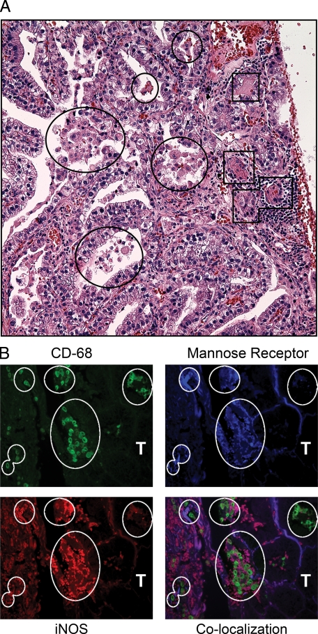
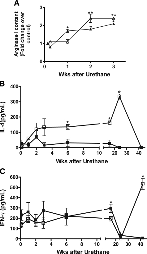
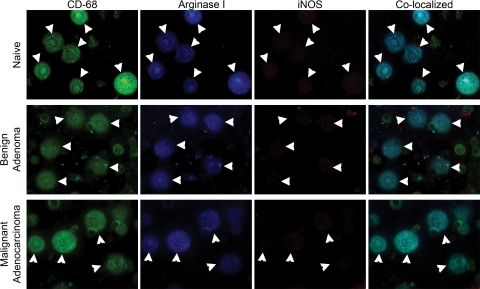
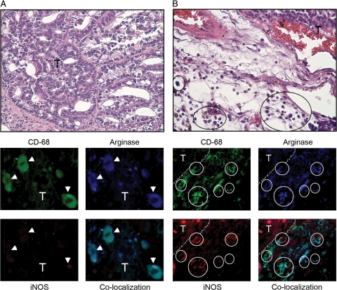
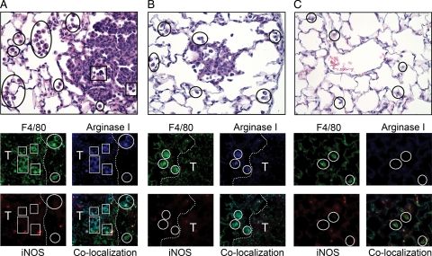
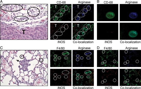
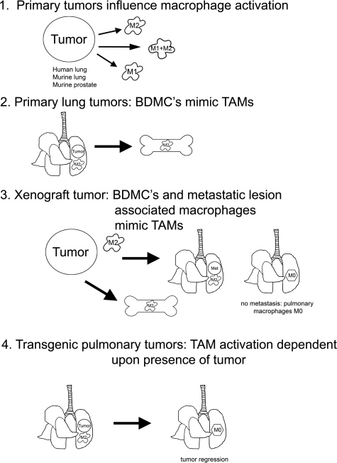
Tumor-associated macrophages (TAMs) encourage and coordinate neoplastic growth. In late stage human lung adenocarcinoma, TAMs exhibited mixed M1 (classical; argI(low)iNOS(high)) and M2 (alternative; argI(high)iNOS(low)) polarization based on arginine metabolism. In several murine cancer models including chemically and genetically-induced primary lung tumors, prostate tumors, colon xenografts, and lung metastases, TAMs expressed argI(high)iNOS(low) early during tumor formation; argI(low)iNOS(high) polarization also occurred during malignancy in some models. In a chemically-induced lung tumor model, macrophages expressed argI(high)iNOS(low) within one week after carcinogen treatment, followed by similar polarization of bone marrow-derived monocytes (BDMCs) a few days later. TAMs surrounding murine prostate tumors also expressed argI(high)iNOS(low) early during tumorigenesis, indicating that this polarization is not unique to neoplastic lungs. In a human colon cancer xenograft model, the primary tumor was surrounded by argI(high)iNOS(low)-expressing TAMs, and BDMCs also expressed argI(high)iNOS(low), but pulmonary macrophages adopted argI(high)iNOS(low) polarization only after tumors metastasized to the lungs. Persistence of tumors is required to maintain TAM polarization. Indeed, in both conditional mutant Kras- and FGF10-driven models of lung cancer, mice expressing the transgene develop lung tumors that regress rapidly when the transgene is silenced. Furthermore, pulmonary macrophages expressed argI(high)iNOS(low) on tumor induction, but then returned to argI(low) iNOS(low) (no polarization) after tumors regressed. Manipulating TAM function or depleting TAMs may provide novel therapeutic strategies for preventing and treating many types of cancer.
Figures
Similar articles
-
Differential polarization of alveolar macrophages and bone marrow-derived monocytes following chemically and pathogen-induced chronic lung inflammation.J Leukoc Biol. 2010 Jul;88(1):159-68. doi: 10.1189/jlb.0609378. Epub 2010 Apr 1. J Leukoc Biol. 2010. PMID: 20360403 Free PMC article.
-
Integrins α5β1 and αvβ3 Differentially Participate in the Recruitment and Reprogramming of Tumor-associated Macrophages in the In Vitro and In Vivo Models of Breast Tumor.J Immunol. 2024 Nov 15;213(10):1553-1568. doi: 10.4049/jimmunol.2400180. J Immunol. 2024. PMID: 39330703
-
Oct4 promotes M2 macrophage polarization through upregulation of macrophage colony-stimulating factor in lung cancer.J Hematol Oncol. 2020 Jun 1;13(1):62. doi: 10.1186/s13045-020-00887-1. J Hematol Oncol. 2020. PMID: 32487125 Free PMC article.
-
A narrative review of tumor-associated macrophages in lung cancer: regulation of macrophage polarization and therapeutic implications.Transl Lung Cancer Res. 2021 Apr;10(4):1889-1916. doi: 10.21037/tlcr-20-1241. Transl Lung Cancer Res. 2021. PMID: 34012800 Free PMC article. Review.
-
The Impact of the Tumor Microenvironment on Macrophage Polarization in Cancer Metastatic Progression.Int J Mol Sci. 2021 Jun 18;22(12):6560. doi: 10.3390/ijms22126560. Int J Mol Sci. 2021. PMID: 34207286 Free PMC article. Review.
Cited by
-
Redox control of inflammation in macrophages.Antioxid Redox Signal. 2013 Aug 20;19(6):595-637. doi: 10.1089/ars.2012.4785. Epub 2013 Mar 6. Antioxid Redox Signal. 2013. PMID: 23311665 Free PMC article. Review.
-
High MUC2 expression in ovarian cancer is inversely associated with the M1/M2 ratio of tumor-associated macrophages and patient survival time.PLoS One. 2013 Dec 6;8(12):e79769. doi: 10.1371/journal.pone.0079769. eCollection 2013. PLoS One. 2013. PMID: 24324582 Free PMC article.
-
CDH3 promotes the progression of lung adenocarcinoma through driving epithelial-mesenchymal transition progress.J Transl Med. 2025 Aug 8;23(1):887. doi: 10.1186/s12967-025-06750-6. J Transl Med. 2025. PMID: 40781712 Free PMC article.
-
Complement and macrophage crosstalk during process of angiogenesis in tumor progression.J Biomed Sci. 2015 Jul 22;22(1):58. doi: 10.1186/s12929-015-0151-1. J Biomed Sci. 2015. PMID: 26198107 Free PMC article. Review.
-
Cinobufagin inhibits M2‑like tumor‑associated macrophage polarization to attenuate the invasion and migration of lung cancer cells.Int J Oncol. 2024 Nov;65(5):102. doi: 10.3892/ijo.2024.5690. Epub 2024 Sep 20. Int J Oncol. 2024. PMID: 39301639 Free PMC article.
References
-
- Nathan C. Points of control in inflammation. Nature. 2002;420:846–852. - PubMed
-
- Geissmann F, Jung S, Littman DR. Blood monocytes consist of two principal subsets with distinct migratory properties. Immunity. 2003;19:71–82. - PubMed
-
- Malkinson AM. Role of inflammation in mouse lung tumorigenesis: a review. Exp Lung Res. 2005;31:57–82. - PubMed
-
- Balkwill F, Mantovani A. Inflammation and cancer: back to Virchow? Lancet. 2001;357:539–545. - PubMed
Publication types
MeSH terms
Substances
Grants and funding
LinkOut - more resources
Full Text Sources
Other Literature Sources
Molecular Biology Databases
Miscellaneous
