. 2010 Jul;30(7):1378-88.
doi: 10.1161/ATVBAHA.109.200428. Epub 2010 Apr 29.

Targeted Vezf1-null mutation impairs vascular structure formation during embryonic stem cell differentiation

Affiliations

Targeted Vezf1-null mutation impairs vascular structure formation during embryonic stem cell differentiation

Zhongmin Zou et al. Arterioscler Thromb Vasc Biol. 2010 Jul.

Abstract

Objective: Vezf1 encodes an early zinc finger transcription factor that is essential for normal vascular development and functions in a dose-dependent manner. Here, we investigated the role of Vezf1 during processes of endothelial cell differentiation and maturation by studying mutant Vezf1 embryonic stem (ES) cells using the in vitro embryoid body differentiation model and the in vivo teratocarcinoma model.

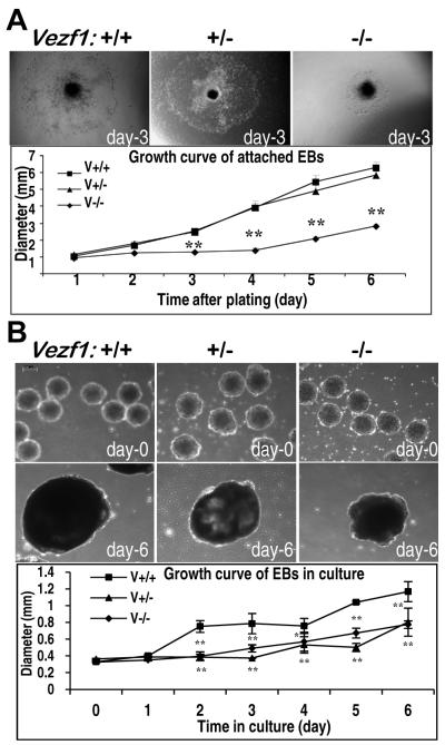
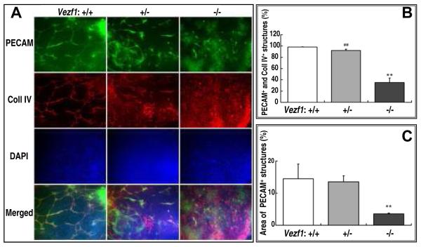
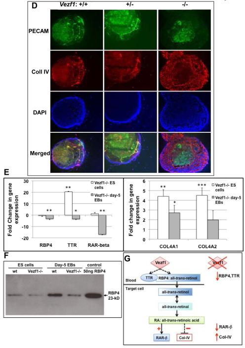
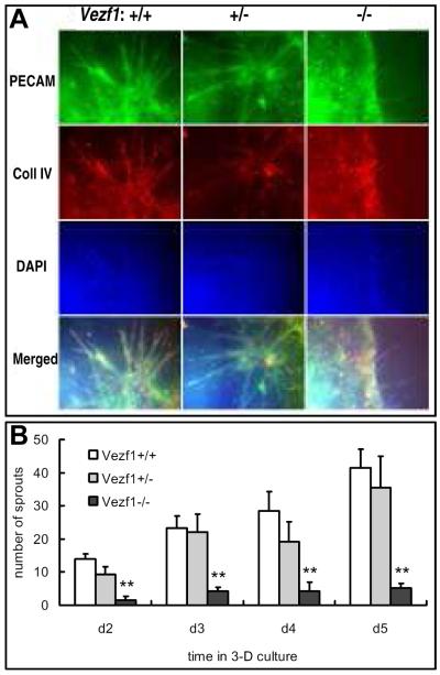
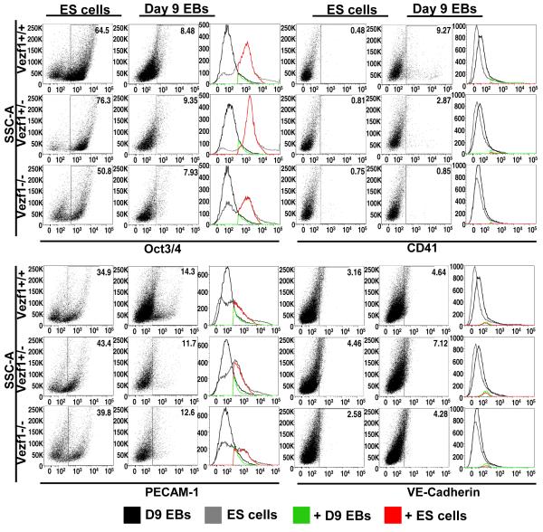
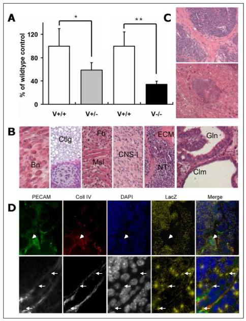
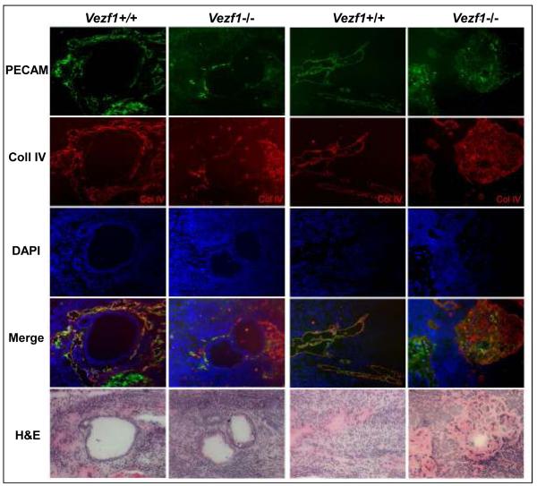
Methods and results: Vezf1-/- ES cell-derived embryoid bodies failed to form a well-organized vascular network and showed dramatic vascular sprouting defects. Our results indicate that the retinol pathway is an important mediator of Vezf1 function and that loss of Vezf1 results in reduced retinol/vitamin A signaling and aberrant extracellular matrix (ECM) formation. Unexpectedly, we also uncovered defects during in vitro differentiation of Vezf1-/- ES cells along hematopoietic cell lineages. Vezf1-/- ES cell-derived teratocarcinomas were able to spontaneously differentiate into cell types of all 3 germ layers. However, histological and immunohistochemical examination of these tumors showed decreased cell proliferation, delayed differentiation, and large foci of cells with extensive deposition of ECM. Embryoid bodies and teratocarcinomas derived from heterozygous ES cells displayed an intermediate phenotype.

Conclusions: Together, these results suggest that Vezf1 is involved in early differentiation processes of the vasculature by regulating cell differentiation, proliferation, and ECM distribution and deposition.


Figures

Figure 1
Figure 1. Growth characteristics of wild-type and Vezf1 mutant embryoid bodies
(A) Growth in 2D attachment culture. EBs were grown for 3 days in suspension culture and switched to 2D attachment culture. EBs attached over night to gelatin-coated plates, and differentiating cells grew out from the center to the periphery. Upper panels: Morphology of representative EBs derived from wt (Vezf1+/+), Vezf1+/−, and Vezf1−/− ES cells, respectively, at day-3 after attachment. Images were taken under an inverted Olympus IX70 microscope. The graph shows growth of EB between day-1 and day-6 of 2D attachment culture. Diameters of EBs (n=14 for each genotype) were measured in 24-hour intervals using Image-Pro Plus software. The experiment was repeated three times, and the graph depicts the results from one representative experiment. **, P<0.01 comparison between wt vs. Vezf1+/− and wt vs. Vezf1+/+ EBs were performed by the student’s t-test. (B) Growth in suspension culture. Upper panels: Representative images of wt, Vezf1+/−, and Vezf1−/− EBs, respectively, at day-0 and day-6 in suspension culture. Images were taken under a Leica DMIL inverted microscope. Bottom panel: Diameters of EBs in suspension culture were measured from day-0 to day-6 using Leica Application Suite (n=5 for each genotype). Comparison between groups were performed by Two-way ANOVA with Bonferroni post tests. *P<0.01, **P<0.001 vs. Vezf1+/− and Vezf1−/− EBs. Data shown as mean ±SEM.
Figure 2
Figure 2. Aberrant vascular structure formation and collagen IV deposition in mutant EBs
(A) EBs in 2D culture Shown are representative images of day-9 EBs derived from wt (Vezf1+/+), Vezf1+/− and Vezf1−/− ES after fixation and immunostaining for PECAM-1 and type IV collagen. Cell nuclei were counter-stained with DAPI. Bottom row depicts merged images from PECAM-1, collagen IV and DAPI staining. Note the extensive vascular network in wt EBs. In contrast, vascular structures did not form properly in Vezf1−/− EBs. Vezf1+/− EBs displayed an intermediate phenotype. Images were taken under a Zeiss Axioplan 2 IE MOT using a 20x objective. (B) Quantitative analysis of vascular coverage. The vasculature as shown by PECAM-1+ staining was analyzed for area density and expressed as percentage of the PECAM-1+ area. At least three independent measurements were performed in each group. Comparison of Vezf1−/− vs. Vezf1+/− and Vezf1+/+ groups were performed by student’s t-test; **, P<0.01. (C) Quantitative analysis of co-expression of PECAM-1 and collagen IV. Shown is the percentage of collagen IV+ areas that was adjacent to or overlapped with PECAM-1+ areas. Comparisons between two groups were done using student’s t-test. **, P<0.01 Vezf1−/− vs. Vezf1+/+; ##, P<0.01 Vezf1−/− vs. Vezf1+/−. (D) Suspension culture EBs. Sections of EBs grown for 9 days in suspension culture were immunostained for PECAM-1 and collagen IV. Cell nuclei were counter-stained with DAPI. Bottom row depicts merged images from PECAM-1, collagen IV and DAPI staining. Well-developed PECAM-1 and collagen IV positive vascular structures were visible in wt and, to a lesser degree, in Vezf1+/− EBs. In Vezf1−/− EBs, PECAM-1 positive sheets of cells were visible in the center, but these failed to form cell cords and they did not co-stain with collagen IV. (E) Expression of retinol pathway genes Rbp4, Ttr, and RAR-β, and collagen 4a1 and 4a2 in ES cells and embryoid bodies. Gene expression levels were determined by quantitative RT-PCR and normalized to β-actin. Shown are changes in expression in Vezf1−/− vs. wt ES cells and day-5 EBs. Comparisons between wt and Vezf1−/− ES cells and EBs were performed by student’s t-test (n=3 for each genotype). (F) Detection of secreted RBP4 protein by western blot analysis. Representative western blot using conditioned media (50μg protein) from wt and Vezf1−/− ES cells and day-5 EBs. 50 ng of purified 23-kD RBP4 protein was loaded as a control. RBP4 levels are reduced in Vezf1−/− EBs and, to a lesser extent, in Vezf1−/− ES cells. (G) Model proposing how Vezf1 regulates the retinol/Vitamin A pathway. Left: Vezf1 activates expression of the all-trans retinol carrier Rbp4 and transthyretin, directly or indirectly. TTR binds to RBP4:all-trans retinol, preventing its renal clearance and resulting in increased cellular uptake. Retinol is converted to retinal and retinoic acid, which directly transactivates expression of its receptor RAR-β and indirectly represses that of Collagen IV. Right: Loss of Vezf1 function in EBs results in deregulation of the retinol/Vitamin A pathways with reduced expression of Rbp4, Ttr and RAR-β, and accumulation of collagen IV.
Figure 3
Figure 3. Vascular sprout formation in EBs cultured in collagen matrix
(A) EBs were cultured for 12 days in suspension culture, followed by 5-day culture in collagen type I gel drops. Endothelial cell sprouts were visualized by whole-mount staining of embedded EBs for PECAM-1 and type IV collagen. Cell nuclei were counter-stained with DAPI. Bottom row depicts merged images from PECAM-1, collagen IV and DAPI staining. Sprout formation was significantly impaired in Vezf1−/− EBs when compared to wt and Vezf1+/− EBs. (B) Quantitative analysis of vascular sprout formation. Individual vascular sprouts from EBs, as indicated by PECAM-1+ staining in corresponding images, were counted at day 2, 3, 4 and 5 in collagen drops (n=10 from each genotype). Comparison of Vezf1−/− vs. Vezf1+/− and Vezf1+/+ groups were performed by student’s t-test; **, P<0.01.
Figure 4
Figure 4. Analysis of Oct3/4, CD41, PECAM-1, and VE-Cadherin gene expression in undifferentiated ES cells and differentiated cells from day-9 EBs
Wild-type, Vezf1+/−, and Vezf1−/− ES cells (106 cells each) and dissociated cells from day-9 suspension culture EBs (550, 880, and 3,300 EBs from wt, Vezf1+/−, and Vezf1−/− EBs, respectively) were analyzed by flow cytometry for the expression of Oct3/4, CD41, PECAM-1 and VE-Cadherin. The gates correspond to Oct3/4+, CD41+, PECAM-1+ or VE-Cadherin+ populations. The histograms depict the number of day-9 EB-derived cell population (black), ES cell population (grey), positive cells from day 9 EB cell population (green), and positive cells from the ES cell population (red).
Figure 5
Figure 5. Teratocarcinoma formation in nude mice
(A) Comparison of weight of teratocarcinomas derived from Vezf1+/− and Vezf1−/− ES cells with their wt controls 2 weeks after subcutaneous injection. The weight of each individual teratocarcinoma was expressed as a percentage of the control (100%) co-injected into the same animal. Six mice each were used for Vezf1+/− and Vezf1−/− ES cells. Vezf1+/− and Vezf1−/− teratocarcinomas were significantly smaller than the wt controls. Values are shown as mean±SE. Comparisons between two groups were performed using student’s t-test. * P<0.05; wt vs. Vezf1+/−, ** P<0.01 wt vs. Vezf1−/−. (B) Histology of representative H&E stained sections of Vezf1−/− teratocarcinomas. From left to right: Bn, bone; Ctlg, cartilage; Fb, fibroblasts; Msl, muscle; CNS-l, central nervous system-like tissue; ECM, extracellular matrix; NT, neural tube; Gln, gland; Clm, cilium. (C) Vezf1−/− teratocarcinomas contained an abundance of areas with less differentiated tissues: multi-cell layered neural tube-like structures with more increased cellularity and decreased lumen formation (top), and extensive deposition of extracellular matrix surrounding neural tube structures (top), or within bone-like structures (bottom). (D) Cryosections of Vezf1lacZ/+ ES cell-derived teratocarcinomas were stained for PECAM-1 and collagen IV, and β-galactosidase. Cell nuclei were counterstained with DAPI. To visualize lacZ in the merged pictures, β-galactosidase staining was shown in a pseudocolor (yellow dots). Most of the PECAM-1, collagen IV positive vessels were negative for lacZ (arrow head). A small fraction of blood vessels contained cells that showed β-galactosidase expression (arrow). Top: 20x objective; bottom: 63x objective.
Figure 6
Figure 6. Vascular structure defects in Vezf1−/− teratocarcinomas
Representative sections from 2 wt and 2 contra-lateral Vezf1−/− teratocarcinomas are shown. Sections were immunostained for PECAM-1 and collagen IV, and cell nuclei were counterstained with DAPI. Merged images are from single PECAM-1, collagen IV and DAPI images. Bottom row: close-by H&E stained section. Note the absence of well-formed vascular structures around glandular epithelial structures in Vezf1−/−, but not wt teratocarcinomas (left 2 columns). Mesenchyme-rich areas in wt teratocarcinomas contained PECAM-1, collagen IV positive vessels. In contrast, Vezf1−/− teratocarcinomas contained large mesenchymal foci with high collagen IV deposition and much lower PECAM-1 expression. These areas did not contain recognizable vascular structures (right 2 columns).

Similar articles

Cited by

References

    1. Carmeliet P. Mechanisms of angiogenesis and arteriogenesis. Nat Med. 2000;6:389–395. - PubMed
    1. Risau W. Mechanisms of angiogenesis. Nature. 1997;386:671–674. - PubMed
    1. Carmeliet P, Jain RK. Angiogenesis in cancer and other diseases. Nature. 2000;407:249–257. - PubMed
    1. Carmeliet P. Angiogenesis in life, disease and medicine. Nature. 2005;438:932–936. - PubMed
    1. Jain RK. Molecular regulation of vessel maturation. Nat Med. 2003;9:685–693. - PubMed

Publication types

MeSH terms