. 2010 Jun;137(11):1787-98.
doi: 10.1242/dev.050799. Epub 2010 Apr 28.

The Puf RNA-binding proteins FBF-1 and FBF-2 inhibit the expression of synaptonemal complex proteins in germline stem cells

Affiliations

The Puf RNA-binding proteins FBF-1 and FBF-2 inhibit the expression of synaptonemal complex proteins in germline stem cells

Christopher Merritt et al. Development. 2010 Jun.

Abstract

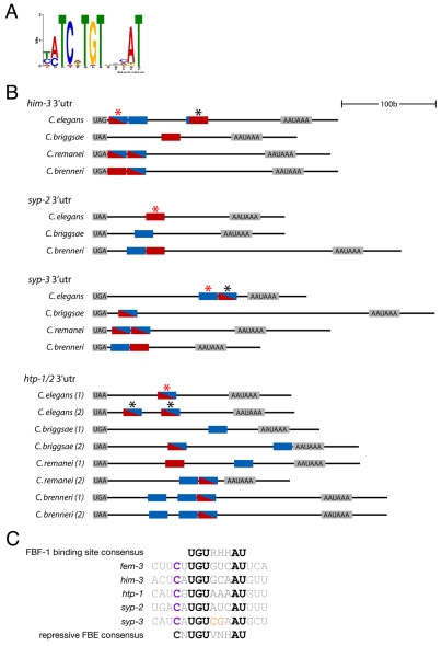
FBF-1 and FBF-2 (collectively FBF) are two nearly identical Puf-domain RNA-binding proteins that regulate the switch from mitosis to meiosis in the C. elegans germline. In germline stem cells, FBF prevents premature meiotic entry by inhibiting the expression of meiotic regulators, such as the RNA-binding protein GLD-1. Here, we demonstrate that FBF also directly inhibits the expression of structural components of meiotic chromosomes. HIM-3, HTP-1, HTP-2, SYP-2 and SYP-3 are components of the synaptonemal complex (SC) that forms between homologous chromosomes during meiotic prophase. In wild-type germlines, the five SC proteins are expressed shortly before meiotic entry. This pattern depends on FBF binding sites in the 3' UTRs of the SC mRNAs. In the absence of FBF or the FBF binding sites, SC proteins are expressed precociously in germline stem cells and their precursors. SC proteins aggregate and SC formation fails at meiotic entry. Precocious SC protein expression is observed even when meiotic entry is delayed in fbf mutants by reducing GLD-1. We propose that parallel regulation by FBF ensures that in wild-type gonads, meiotic entry is coordinated with just-in-time synthesis of synaptonemal proteins.


Figures

Fig. 1.
Fig. 1.
Summary of 3′ UTR fusions examined in this study. (A) The distal end of the C. elegans adult gonadal tube. Development proceeds from distal (left) to proximal (right). The transition zone where germ cells initiate meiotic prophase is characterized by a distinct crescent chromosomal morphology and is marked by a vertical dashed line in this and all other figures. The proximal half of the `mitotic zone' includes cells that have initiated meiotic S phase (reviewed by Hubbard, 2007). (B) Expression patterns of the 3′ UTR reporters examined in this study. Genes are arranged by functional class. Dark gray indicates the strongest domain(s) of GFP::histone H2B expression, light gray indicates weaker domain(s) of GFP expression, and white indicates no GFP expression. (C) The design of the 3′ UTR fusions. The pie-1 promoter is permissive for expression in all germ cells (Merritt et al., 2008). GFP::histone H2B provides the protein reporter. (D) Expression of selected 3′ UTR reporters in the distal end of the gonad. The gonad is outlined.
Fig. 2.
Fig. 2.
Conserved motifs in the 3′ UTRs of synaptonemal complex (SC) genes and their homologs. (A) Conserved motif found in the SC 3′ UTRs by MEME (see Materials and methods). (B) Shown are sites matching the MEME motif (blue boxes, UCNUGUNNNAU), sites matching the in vitro-defined preferred FBF-1 binding site (red boxes, UGU A/G A/C/U A/C/U AU) (Bernstein et al., 2005), and sites matching both (red and blue boxes) in SC 3′ UTRs from C. elegans, C. briggsae, C. remanei and C. brenneri. Asterisks denote sites mutated (from UGUNNNAU to acaNNNAU) in reporters shown in Fig. 3. Red asterisks denote sites unambiguously required for repression by FBF. Sites in him-3 were mutated singly (data not shown) and in combination (see Fig. 3); mutation in the first site was sufficient for full derepression (data not shown). The second site in syp-3 was mutated singly and had no effect (data not shown), whereas the first site in syp-3 was mutated in combination with the second site and found to cause derepression (see Fig. 3). (C) Alignment of sites required for repression in the SC 3′ UTRs (red asterisks in B) and in the fem-3 3′ UTR (Ahringer and Kimble, 1991). A 5′ cytosine is conserved in all (purple). The syp-3 site has two bases (orange) that do not fit the in vitro-defined FBF-1 binding site. Note that all sites, except for syp-2, also have a uracil before the conserved 5′ cytosine. The repressive FBF-response element (FBE) consensus is based on all the sequences shown here (see Discussion).
Fig. 3.
Fig. 3.
FBF and predicted FBF binding sites are required for downregulation of SC 3′ UTR reporters in the mitotic zone. (A) Fluorescence photomicrographs of distal arms from C. elegans adult hermaphrodites expressing the indicated 3′ UTR fusions, and fed bacteria containing either the empty feeding vector (L4440), the fbf-1 and fbf-2 feeding vectors [fbf-1/2(RNAi)], or no vector (FBE mutant). FBE mutants are the 3′ UTR reporters mutated at the predicted FBF sites shown in Fig. 2. See Table S2 and Fig. S2 in the supplementary material for numbers and additional 3′ UTR fusions examined. (B) Fluorescence photomicrographs of L2 gonadal arms expressing the indicated 3′ UTR reporters and treated as in A. In the case of him-3, syp-2, syp-3 and puf-5, the 3′ UTR reporter fusions were examined in hermaphrodites heterozygous (fbf-1/2/mIn1) or homozygous (fbf-1/2) for mutations in fbf-1 and fbf-2 [fbf-1(ok91);fbf-2(q704)]. (C) Fluorescence photomicrographs of L4 gonads expressing the indicated 3′ UTR fusions in fbf-1(ok91), fbf-2(q738) and fbf-1(ok91);fbf-2(q704) mutant larvae. Note that in all cases, moderate derepression of the transgenes is observed in the fbf-1 single mutant but not in the fbf-2 single mutant, suggesting that FBF-1 is only partially redundant with FBF-2 in this assay.
Fig. 4.
Fig. 4.
FBF-1 and FBF-2 interact with endogenous SC mRNAs in vivo. (A) Extracts from worms expressing GFP::FBF-1, GFP::FBF-2 and GFP::TBB-2 (β-tubulin) were immunoprecipitated with either a GFP antibody or IgG. Immunoprecipitates were blotted for GFP or α-tubulin. Input is 1/16 of the immunoprecipitation (IP). (B) Enrichment of transcripts in GFP versus IgG immunoprecipitates. Values are normalized to actin mRNA enrichment to correct for non-specific binding of RNAs during the GFP IP. gld-1 mRNA was enriched 74- and 42-fold in GFP::FBF-1 and GFP::FBF-2 IPs, respectively. Error bars represent s.e.m. FBF-1 binding sites are UGU A/G A/C/U A/C/U AU. Repressive FBE sites are CNUGU A/G/C N A/C/U AU (as in Fig. 2).
Fig. 5.
Fig. 5.
Precocious expression of SC proteins in C. elegans fbf-1;fbf-2 mutants. (A) Fluorescence photomicrographs of L4 gonads. HIM-3 and HTP-1/2 expression was assessed by antibody staining (anti-HTP recognizes both HTP-1 and HTP-2) in hermaphrodites heterozygous (fbf-1/2/mIn1) or homozygous (fbf-1/2) for mutations in fbf-1 and fbf-2. The HIM-3 antibody cross-reacts with mitotic spindles and other tubulin-rich structures (data not shown) and therefore some background cytoplasmic staining is visible in distal cells in the fbf-1/2/mIn1 control. SYP-2 and SYP-3 expression was examined using GFP fusions (containing the full syp-2 and syp-3 loci) in live animals that were fed bacteria containing either the empty feeding vector (L4440) or feeding vectors for fbf-1 and fbf-2. Arrows point to aggregates. (B) Fluorescence photomicrographs of L2 gonadal arms expressing the indicated GFP fusions. At this stage, all germ cells are proliferating. The GFP::SC fusions are not expressed in control gonads, but are expressed in all cells in gonads lacking fbf-1 and fbf-2. (C) Fluorescence photomicrographs of embryos expressing the indicated GFP fusions driven by the inducible heat-shock promoter hsp16-41. The SC fusions form aggregates not seen with the GFP::histone H2B fusion.
Fig. 6.
Fig. 6.
Defective synaptonemal complexes in C. elegans fbf-1;fbf-2 mutants. (A) Fluorescence photomicrographs of pachytene-stage germ cells in early and middle L4 gonads stained with anti-HTP-1/2 or expressing GFP::SYP-3, comparing hermaphrodites heterozygous (fbf-1/2/mIn1) or homozygous (fbf-1/2) for mutations in fbf-1 and fbf-2 or wild-type hermaphrodites treated with control feeding vector (L4440) or fbf-1/2 RNAi. Arrows point to representative large foci not seen in controls. (B) Distal gonads expressing GFP::HIM-3 in adult hermaphrodites of the indicated genotypes. All germ cells undergo spermatogenesis in fbf-1;fbf-2 homozygotes grown at 20°C (GFP::HIM-3 is downregulated during spermatogenesis so no fluorescence is visible). gld-1 RNAi or growth at 25°C restores a mitotic zone in fbf-1;fbf-2 adults, but this zone still misexpresses GFP::HIM-3.

Similar articles

Cited by

References

    1. Ahringer J., Kimble J. (1991). Control of the sperm-oocyte switch in Caenorhabditis elegans hermaphrodites by the fem-3 3′ untranslated region. Nature 349, 346-348 - PubMed
    1. Anderson E. L., Baltus A. E., Roepers-Gajadien H. L., Hassold T. J., de Rooij D. G., van Pelt A. M., Page D. C. (2008). Stra8 and its inducer, retinoic acid, regulate meiotic initiation in both spermatogenesis and oogenesis in mice. Proc. Natl. Acad. Sci. USA 105, 14976-14980 - PMC - PubMed
    1. Bailey T. L., Elkan C. (1994). Fitting a mixture model by expectation maximization to discover motifs in biopolymers. Proc. Int. Conf. Intell. Syst. Mol. Biol. 2, 28-36 - PubMed
    1. Bernstein D., Hook B., Hajarnavis A., Opperman L., Wickens M. (2005). Binding specificity and mRNA targets of a C. elegans PUF protein, FBF-1. RNA 11, 447-458 - PMC - PubMed
    1. Bhalla N., Wynne D. J., Jantsch V., Dernburg A. F. (2008). ZHP-3 acts at crossovers to couple meiotic recombination with synaptonemal complex disassembly and bivalent formation in C. elegans. PLoS Genet. 4, e1000235 - PMC - PubMed

Publication types

MeSH terms