. 2010 Jun;137(11):1833-42.
doi: 10.1242/dev.047647. Epub 2010 Apr 28.

PAR-3 mediates the initial clustering and apical localization of junction and polarity proteins during C. elegans intestinal epithelial cell polarization

Affiliations

PAR-3 mediates the initial clustering and apical localization of junction and polarity proteins during C. elegans intestinal epithelial cell polarization

Annita Achilleos et al. Development. 2010 Jun.

Abstract

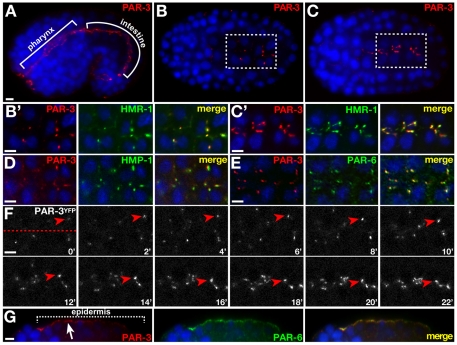
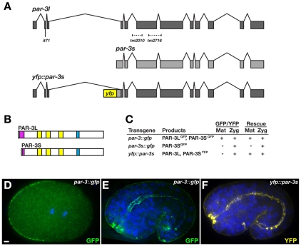
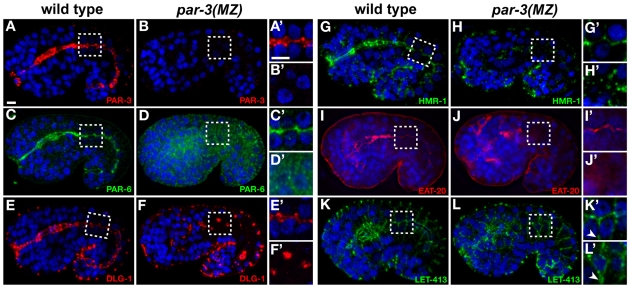
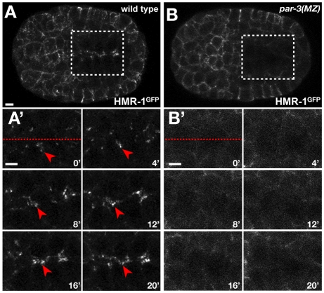
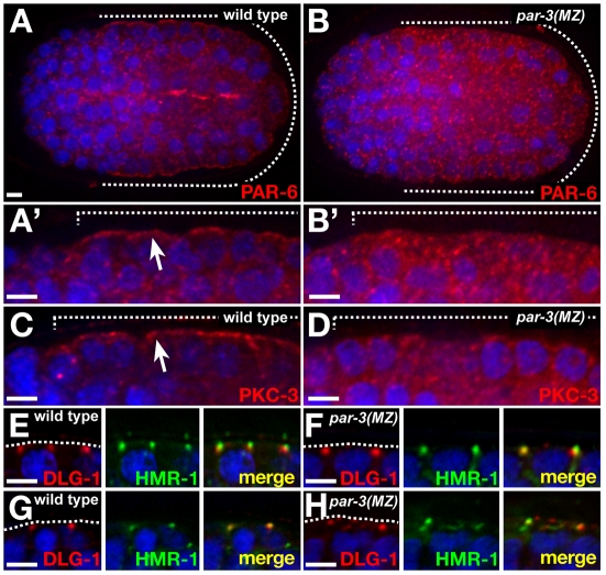
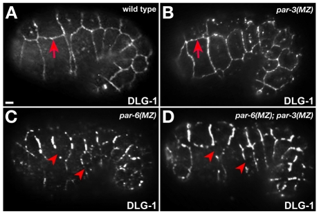
The apicobasal polarity of epithelial cells is critical for organ morphogenesis and function, and loss of polarity can promote tumorigenesis. Most epithelial cells form when precursor cells receive a polarization cue, develop distinct apical and basolateral domains and assemble junctions near their apical surface. The scaffolding protein PAR-3 regulates epithelial cell polarity, but its cellular role in the transition from precursor cell to polarized epithelial cell has not been determined in vivo. Here, we use a targeted protein-degradation strategy to remove PAR-3 from C. elegans embryos and examine its cellular role as intestinal precursor cells become polarized epithelial cells. At initial stages of polarization, PAR-3 accumulates in cortical foci that contain E-cadherin, other adherens junction proteins, and the polarity proteins PAR-6 and PKC-3. Using live imaging, we show that PAR-3 foci move apically and cluster, and that PAR-3 is required to assemble E-cadherin into foci and for foci to accumulate at the apical surface. We propose that PAR-3 facilitates polarization by promoting the initial clustering of junction and polarity proteins that then travel and accumulate apically. Unexpectedly, superficial epidermal cells form apical junctions in the absence of PAR-3, and we show that PAR-6 has a PAR-3-independent role in these cells to promote apical junction maturation. These findings indicate that PAR-3 and PAR-6 function sequentially to position and mature apical junctions, and that the requirement for PAR-3 can vary in different types of epithelial cells.


Figures

Fig. 1.
Fig. 1.
PAR-3 localization within polarizing epithelial cells. DNA is blue (in this and all subsequent figures). Anterior is left. C. elegans embryos are ∼50 μm. (A-C′) PAR-3 in polarized intestinal and pharyngeal cells (A), in intestinal precursor cells (IPCs) that are beginning to polarize (B), or during polarization (C). The boxed regions in B,C are shown in B′,C′ and show colocalization of PAR-3 with HMR-1. (D,E) PAR-3 colocalization with HMP-1 (D) and PAR-6 (E) in IPCs at the onset of polarization. (F) Stills from Movie 2 of IPCs expressing PAR-3YFP (see Movie 2 in the supplementary material). Dashed line indicates the future apical surface. A focus of PAR-3YFP (arrowhead) is shown over time (minutes). (G) Lateral view of polarizing epidermis (bracketed region) showing apical PAR-3 (arrow) and PAR-6. Scale bars: 2.5 μm.
Fig. 2.
Fig. 2.
Isoforms of C. elegans par-3. (A) par-3l, par-3s and yfp::par-3s. Exons (rectangles), introns (chevrons) and mutations are indicated. (B) Predicted PAR-3L and PAR-3S products showing the oligomerization domain (magenta), PDZ domains (yellow) and aPKC-binding domain (cyan). (C) Expression and rescuing activity of par-3 transgenes. (D,E) Expression of par-3::gfp in one-cell (D) and 1.5-fold stage (E) embryos. (F) Expression of yfp::par-3s in 1.5-fold stage embryo. Scale bar: 2.5 μm.
Fig. 3.
Fig. 3.
Morphogenesis in par-3(MZ) embryos. (A,B) DIC time-lapse stills of wild-type and par-3(MZ) mutant C. elegans embryos at 1.5-fold stage. The pharynx (outlined) does not extend anteriorly in the par-3(MZ) embryo and extruded cells (arrowheads) are visible. (C) Elongation rate to 1.5-fold stage. Time is minutes from completion of ventral enclosure to 1.5-fold stage. Error bars indicate s.d.; *, P<0.003 (Student's t-test). Scale bar: 2.5 μm.
Fig. 4.
Fig. 4.
Junction and polarity proteins in par-3(MZ) embryos. (A-L′) Wild-type and par-3(MZ) C. elegans embryos at 1.25- to 1.5-fold stage, immunostained as indicated. The boxed regions are shown at higher magnification to the right; the contrast in some of these has been increased to highlight local staining. Arrowheads indicate LET-413 at lateral surfaces. Scale bars: 2.5 μm.
Fig. 5.
Fig. 5.
HMR-1GFP localization during epithelial cell polarization. (A-B′) Stills from movies of wild-type (A, from Movie 5) and par-3(MZ) (B, from Movie 6) C. elegans embryos expressing HMR-1GFP (see Movies 5, 6 in the supplementary material). The boxed regions in A,B are shown in A′,B′ in a time-lapse sequence covering the preceding 20 minutes. 0′ represents the time at which HMR-1GFP is first detected in foci in wild-type IPCs. Dashed lines in A′,B′ denote the future apical surface. Scale bars: 2.5 μm.
Fig. 6.
Fig. 6.
Epidermal epithelial cells in par-3(MZ) embryos. (A-B′) PAR-6 staining in the epidermis (underlying the dashed lines). The epidermis is shown at higher magnification in A′,B′. (C,D) PKC-3 staining in the epidermis. (E-H) Epidermal cells showing DLG-1 and HMR-1 in apical junctions at comma stage (E,F) or 1.5-fold stage (G,H), when junction proteins become more dispersed in par-3(MZ) than in wild-type C. elegans embryos. Scale bars: 2.5 μm.
Fig. 7.
Fig. 7.
Junction maturation in par-6(MZ); par-3(MZ) double-mutant embryos. (A-D) Superficial views of 1.25- to 1.5-fold stage embryos stained for DLG-1. Junctions are continuous (arrows) in wild-type and par-3(MZ) C. elegans embryos, but thick and fragmented (arrowheads) in par-6(MZ) and par-6(MZ); par-3(MZ) embryos. Scale bar: 2.5 μm.

References

    1. Anderson D. C., Gill J. S., Cinalli R. M., Nance J. (2008). Polarization of the C. elegans embryo by RhoGAP-mediated exclusion of PAR-6 from cell contacts. Science 320, 1771-1774 - PMC - PubMed
    1. Aono S., Legouis R., Hoose W. A., Kemphues K. J. (2004). PAR-3 is required for epithelial cell polarity in the distal spermatheca of C. elegans. Development 131, 2865-2874 - PubMed
    1. Bilder D. (2004). Epithelial polarity and proliferation control: links from the Drosophila neoplastic tumor suppressors. Genes Dev. 18, 1909-1925 - PubMed
    1. Bossinger O., Klebes A., Segbert C., Theres C., Knust E. (2001). Zonula adherens formation in Caenorhabditis elegans requires dlg-1, the homologue of the Drosophila gene discs large. Dev. Biol. 230, 29-42 - PubMed
    1. Bossinger O., Fukushige T., Claeys M., Borgonie G., McGhee J. D. (2004). The apical disposition of the Caenorhabditis elegans intestinal terminal web is maintained by LET-413. Dev. Biol. 268, 448-456 - PubMed

Publication types

MeSH terms