. 2009 Feb 18;1(1):2-17.
doi: 10.1111/j.1757-1707.2009.01004.x.

Enzymatic deconstruction of xylan for biofuel production

Affiliations

Enzymatic deconstruction of xylan for biofuel production

Dylan Dodd et al. Glob Change Biol Bioenergy. .

Abstract

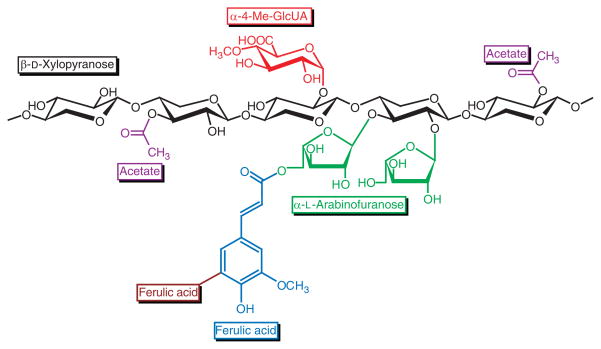
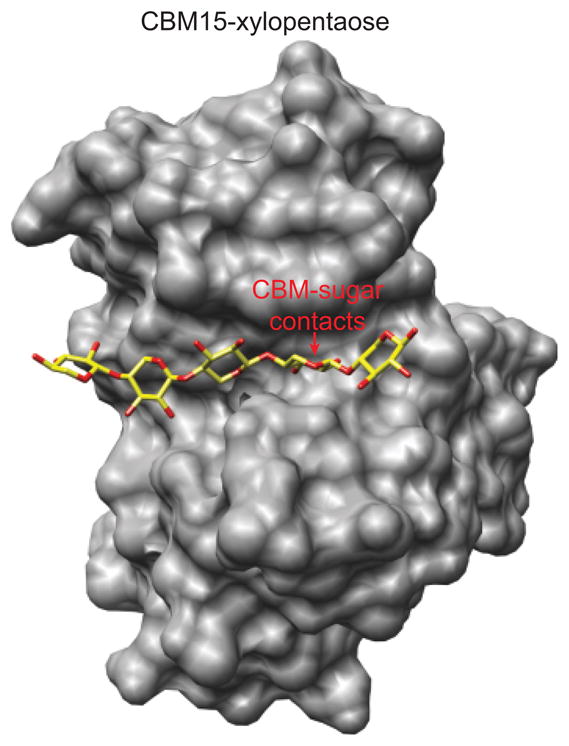
The combustion of fossil-derived fuels has a significant impact on atmospheric carbon dioxide (CO(2)) levels and correspondingly is an important contributor to anthropogenic global climate change. Plants have evolved photosynthetic mechanisms in which solar energy is used to fix CO(2) into carbohydrates. Thus, combustion of biofuels, derived from plant biomass, can be considered a potentially carbon neutral process. One of the major limitations for efficient conversion of plant biomass to biofuels is the recalcitrant nature of the plant cell wall, which is composed mostly of lignocellulosic materials (lignin, cellulose, and hemicellulose). The heteropolymer xylan represents the most abundant hemicellulosic polysaccharide and is composed primarily of xylose, arabinose, and glucuronic acid. Microbes have evolved a plethora of enzymatic strategies for hydrolyzing xylan into its constituent sugars for subsequent fermentation to biofuels. Therefore, microorganisms are considered an important source of biocatalysts in the emerging biofuel industry. To produce an optimized enzymatic cocktail for xylan deconstruction, it will be valuable to gain insight at the molecular level of the chemical linkages and the mechanisms by which these enzymes recognize their substrates and catalyze their reactions. Recent advances in genomics, proteomics, and structural biology have revolutionized our understanding of the microbial xylanolytic enzymes. This review focuses on current understanding of the molecular basis for substrate specificity and catalysis by enzymes involved in xylan deconstruction.


Figures

Fig. 1
Fig. 1
Compositional analysis of the Alamo cultivar of Switch-grass. As indicated, glucans are predominantly composed of cellulose. Hemicellulosic components include galactan, mannan, xylan, arabinan, and uronic acids, although xylan represents the most abundant hemicellulosic polymer (Vogel, 2008). These data were obtained from the US DOE Biomass Feedstock Composition and Property Database (http://www.afdc.energy.gov/biomass/progs/search1.cgi).
Fig. 2
Fig. 2
General structure showing the various linkages found in a variety of xylans isolated from plant cell walls. As described in the text, xylans isolated from different sources may not possess all of the linkages shown. Ferulic acid may form a di-ferulic acid bridge with ferulic acid residues attached to other arabinoxylan chains. Xylose residues may be di-substituted or mono-substituted with arabinose at the O-2 and O-3 positions.
Fig. 3
Fig. 3
General glycosidase mechanisms for (a) retaining glycosidases and (b) inverting glycosidases. In (a), a deprotonated carboxylic acid nucleophile attacks the anomeric carbon, displacing the attached sugar residue (indicated as R) and forming a covalent enzyme–sugar adduct. Subsequently an activated water molecule displaces the enzymic carboxylic acid resulting in net retention of stereochemical configuration at the anomeric carbon. In (b), an activated water molecule displaces the attached sugar residue resulting in net inversion of stereochemical configuration. In both of these mechanisms, the glycoside leaving group is assisted through protonation by a catalytic acid residue.
Fig. 4
Fig. 4
Diagrammatic representation of the sugar binding sites in glycosidases based on the scheme proposed by Davies et al. (1997).
Fig. 5
Fig. 5
Differences in the products of hydrolysis between GH 10 and 11 endo-xylanases when incubated with substituted xylans (Biely et al., 1997b; Maslen et al., 2007). (a) For GH family 10 enzymes, substitutions on the xylan chain are accommodated at the +1 site, thus these enzymes can release xylo-oligosaccharides in which the terminal nonreducing xylose residue is substituted. (b) For GH family 11 enzymes, substitutions on the xylan chain are not accommodated at the +1 site, thus these enzymes produce xylo-oligosaccharides with substitutions at the penultimate xylose residue. GH, glycoside hydrolase.
Fig. 6
Fig. 6
Structural surface representation of the (a) Neocallimastix patriciarum GH family 11 xylanase in complex with ferulic acid arabinoxylotrisaccharide (PDB accession no. 2VGD) (Vardakou et al., 2008) and the (b) Cellvibrio mixtus GH family 10 xylanase in complex with aldotetraouronic acid (PDB accession no. 1UQZ) (Pell et al., 2004b). For (a), the active site cleft of the GH 11 enzyme excludes the possibility of a substitution at the +1 subsite of the xylose chain, whereas substitutions may be accommodated at the +2 subsite. For (b), the open topology of the active site of the GH 10 enzyme permits the accommodation of a substitution at the +1 subsite. All structural representations in this and subsequent figures were generated with the UCSF Chimera software package (Pettersen et al., 2004). GH, glycoside hydrolase.
Fig. 7
Fig. 7
Structural representations of the (a) Bacillus subtilis GH family 43 arabinoxylan arabinofuranohydrolase (AXH-m2,3) in complex with xylotetraose (PDB accession no. 3C7G) (Vandermarliere et al., 2009) and the (b) Selenomonas ruminantium GH family 43 β-xylosidase (SXA) in complex with 1,3-bis[tris(hydroxymethyl)methylamino]propane (PDB accession no. 3C2U) (Brunzelle et al., 2008). Both enzymes possess two domains, an N-terminal β-propeller domain and a C-terminal mainly β-sheet domain, although the C-terminal domain for SXA is much larger and projects a loop that contacts the active site for the enzyme. GH, glycoside hydrolase.
Fig. 8
Fig. 8
Schematic representation of the GH family 43 β-xylosidase (SXA) from Selenomonas ruminantium indicating the two xylose binding sites in the active site and the projection of extended xylose chains out into solution. GH, glycoside hydrolase.
Fig. 9
Fig. 9
Structural representations of the (a) Vibrio cholerae putative GH family 3 N-acetyl-β-glucosaminidase in complex with N-acetyl-β-glucosaminidase (PDB accession no. 1Y65) and the (b) Hordeum vulgare GH family 3 β-xylosidase in complex with glucose (PDB accession no. 1EX1) (Varghese et al., 1999). As described in the text, acquisition of the second (α/β)6 sandwich domain may have led to the evolution of β-glucosidase activity within this family of proteins. GH, glycoside hydrolase.
Fig. 10
Fig. 10
General mechanism for esterases employing the Ser–His–Asp(Glu) catalytic triad. Analogous to serine proteases, the first set of reactions leads to acylation of the enzyme followed by de-acylation of the enzyme involving attack of the ester linkage by an activated water molecule.
Fig. 11
Fig. 11
Structural surface rendering of the Cellvibrio japonicus family 15 carbohydrate binding module (CBM) in complex with xylopentaose (PDB accession no. 1GNY) (Szabo et al., 2001). The xylopentaose sugar adopts a helical conformation wherein most of the O-2 and O-3 hydroxyl groups point out into solution suggesting that this CBM could accommodate a highly decorated xylan chain. The only exception is the fourth xylose residue which makes hydrogen bond contacts from both the O-2 and O-3 hydroxyl groups to the protein.
Fig. 12
Fig. 12
Schematic outline depicting the functional coordination of xylanolytic enzymes in the deconstruction of xylan for biofuels production. (a) Xylanases, acetyl xylan esterases, and ferulic acid esterases function together to produce short, substituted xylo-oligosaccharides with the concomitant release of ferulic and acetic acid byproducts. (b) Arabinofuranosidases and glucuronidases then liberate arabinose and glucuronic acid from these substituted xylo-oligosaccharides. (c) Xylosidases convert the xylo-oligosaccharides into their constituent xylose sugars. (d) Fermenting microorganisms take up the xylose and arabinose and shuttle them into the pentose phosphate pathway for subsequent fermentation to biofuels.

References

    1. Agbogbo FK, Coward-Kelly G. Cellulosic ethanol production using the naturally occurring xylose-fermenting yeast, Pichia stipitis. Biotechnology Letters. 2008;30:1515–1524. - PubMed
    1. Antal MJ, Jr, Mok WS, Richards GN. Mechanism of formation of 5-(hydroxymethyl)-2-furaldehyde from D-fructose and sucrose. Carbohydrate Research. 1990;199:91–109. - PubMed
    1. Bailey MJ, Biely P, Poutanen K. Interlaboratory testing of methods for assay of xylanase activity. Journal of Biotechnology. 1992;23:257–270.
    1. Bayer EA, Lamed R, Himmel ME. The potential of cellulases and cellulosomes for cellulosic waste management. Current Opinion in Biotechnology. 2007;18:237–245. - PubMed
    1. Berrin JG, Juge N. Factors affecting xylanase functionality in the degradation of arabinoxylans. Biotechnology Letters. 2008;30:1139–1150. - PubMed

LinkOut - more resources