. 2009 Sep;3(3):145-65.
doi: 10.5009/gnl.2009.3.3.145. Epub 2009 Sep 30.

Liver transplantation

Affiliations

Liver transplantation

Deok-Bog Moon et al. Gut Liver. 2009 Sep.

Abstract

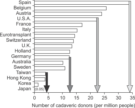
Liver transplantation has become a lifesaving procedure for patients who have chronic end-stage liver disease and acute liver failure. The satisfactory outcome of liver transplantation has led to insufficient supplies of deceased donor organs, particularly in East Asia. Hence, East Asian surgeons are concentrating on developing and performing living-donor liver transplantation (LDLT). This review article describes an update on the present status of liver transplantation, mainly in adults, and highlights some recent developments on indications for transplantation, patient selection, donor and recipient operation between LDLT and deceased-donor liver transplantation (DDLT), immunosuppression, and long-term management of liver transplant recipients. Currently, the same indication criteria that exist for DDLT are applied to LDLT, with technical refinements for LDLT. In highly experienced centers, LDLT for high-scoring (>30 points) Model of End-Stage Liver Disease (MELD) patients and acute-on-chronic liver-failure patients yields comparably good outcomes to DDLT, because timely liver transplantation with good-quality grafting is possible. With increasing numbers of liver transplantations and long-term survivors, specialized attention should be paid to complications that develop in the long term, such as chronic renal failure, hypertension, diabetes mellitus, dyslipidemia, obesity, bone or neurological complications, and development of de novo tumors, which are highly related to the immunosuppressive treatment.

Keywords: Deceased donor liver transplantation; Liver transplantation; Living donor liver transplantation.


Figures

Fig. 1
Fig. 1
Number of deceased donor livers available in various countries in 2000. Reprinted from de Villa VH, Lo CM, Chen CL. Ethics and rationale of living-donor liver transplantation in Asia. Transplantation 2003;75(3 Suppl):S2-S5.
Fig. 2
Fig. 2
Annual number of liver transplantations performed at the Asan Medical Center, Ulsan University College of Medicine. The reduction in the accepted remnant liver volume from >35% to >30% of the donor's total liver volume in 2005 resulted in a decrease in the proportion of dual-graft LDLTs. In 2008 there was a marked increase in organ donations, which was fueled by organ donation from a famous boxer in early 2008, hence reducing the number of LDLTs. LDLT, living donor liver transplantation; DDLT, deceased donor liver transplantation.
Fig. 3
Fig. 3
Comparison of indications for liver transplantation between Korea and the United States. HBV, hepatitis B virus; LC, liver cirrhosis; HCC, hepatocellular carcinoma; FHF, fulminant hepatic failure; HCV, hepatitis C virus; SBC, secondary biliary cirrhosis; PBC, primary biliary cirrhosis; AIH, autoimmune hepatitis; CCC, cholangiocarcinoma; Re-LT, retransplantation; PSC, primary sclerosing cholangitis; BD, bile duct.
Fig. 4
Fig. 4
LDLT with replacement of the inferior vena cava using a Dacron interposition graft for Budd-Chiari syndrome. (A) Preoperative CT scan showing typical findings of Budd-Chiari syndrome: retrohepatic obliteration of the inferior vena cava (multiple arrows), and portal vein stenosis and a large coronary collateral vein (black and white arrowheads, respectively). (B) Intraoperative photograph showing replacement of the inferior vena cava with a Dacron interposition graft between the right atrium and the suprarenal inferior vena cava (white arrowhead), and middle hepatic vein tributaries reconstructed using cryopreserved cadaveric iliac vein grafts. (C) Two-year postoperative CT scan revealing a patent replaced inferior vena cava, right hepatic vein, and portal vein without stenosis (white arrowhead, white arrow, and black arrowhead, respectively). LDLT, living donor liver transplantation.
Fig. 5
Fig. 5
Portal vein plasty with the great saphenous vein and intraoperative stent placement for severe portal vein stenosis. (A) Preoperative CT scan showing severe portal vein stenosis and abundant ascites. (B) The liver graft regenerated well and ascites had disappeared by the 2-month postoperative follow-up, as revealed by this CT scan, after intraoperative portal vein plasty with an autogenous great saphenous vein (GSV, indicated by the separate figure in the small box) and additional stent placement (black arrow).
Fig. 6
Fig. 6
Renoportal anastomosis using a cadaveric fresh iliac vein graft for an obliterated portal vein, and large spontaneous splenorenal shunts. (A) Preoperative CT scan showing obliterated portal vein stenosis and large spontaneous splenorenal shunts draining most of the splanchnic blood flow into the left renal vein (LRV, black arrow). (B) Intraoperative photography showing an interposition cadaveric fresh iliac vein graft (IPG) anastomosed to the left renal vein. The black arrow indicates the interruption suture running between the inferior vena cava (IVC) and the left renal vein in order to prevent portal flow stealing. (C) Patent interposition iliac vein graft between the left renal vein and the grafted portal vein. A well-regenerated liver is visible on the 2-year postoperative follow-up CT scan.
Fig. 7
Fig. 7
CT scan showing changes between before and after liver transplantation. (A) HBV-related acute-on-chronic liver failure patient with huge ascites, altered mentality, and high MELD score (40 points). (B) Good regeneration of dual liver grafts is visible and the ascites have disappeared on this 1-month postoperative follow-up CT scan.
Fig. 8
Fig. 8
Interrelationship between graft volume, hepatic inflow and outflow, and bile-duct anastomosis for determining the technically successful living donor liver transplantation (LDLT).
Fig. 9
Fig. 9
Split liver transplantation to two adult recipients in order to expand the donor pool at Asan Medical Center. This is the first time that this procedure was performed in Korea, in August 2003.
Fig. 10
Fig. 10
Segmental anatomy of the liver using the Couinaud's segments. Reprinted from Brown RS Jr. Live donors in liver transplantation. Gastroenterology 2008;134:1802-1813.
Fig. 11
Fig. 11
Most commonly used graft for adult-to-adult LDLT. (A) Right lobe graft, (B) Left lobe graft.
Fig. 12
Fig. 12
Congestion of the medial sector after procurement of the right lobe with a middle hepatic vein graft from a living liver donor. (A) Preoperative CT scan showing the donor liver and imaginary parenchymal transection line when the right lobe with the middle hepatic vein graft is harvested. (B) CT scan made on the 7th postoperative day, revealing a large area of congestion at the medial sector.
Fig. 13
Fig. 13
A scheme for modified right lobe (MRL) liver grafting. RHV, right hepatic vein; MHV, middle hepatic vein; LHV, left hepatic vein; V5, hepatic venous tributaries from segment 5; V8, hepatic venous tributaries from segment 8.
Fig. 14
Fig. 14
Postoperative follow-up CT scan of the recipient, demonstrating the balanced regeneration of both liver grafts. (A) CT scan taken 5 days after transplantation showing that the second left lobe graft in the right upper abdomen was still small and supported by a tissue expander bag. (B) CT scan made 2 weeks after transplantation, showing the rapid regeneration of both grafts. (C) CT scan made 2 months after transplantation, showing that two regenerated left lobe grafts were in the shape of a normal liver.
Fig. 15
Fig. 15
Two hundred and fifty-eight dual-graft adult-to-adult LDLTs were performed between March 2000 and December 2008 at the Asan Medical Center. *Two recipients received a left lobe from a live donor and a split lateral segment from a deceased donor, respectively, on June 2000 and December 2008, respectively; Right lobe (from wife) and left lobe (from cousin) dual-graft adult-to-adult LDLT for a large-size recipient was performed successfully in April 2001. Lateral section replaced the left lobe in 10 of 47 recipients.
Fig. 16
Fig. 16
Types of graft in 1,739 adult-to-adult LDLTs. MRL, modified right lobe; RL, right lobe; LL, left lobe; S1, caudate lobe; ERL, extended right lobe; RPS, right posterior sector; E-LLS, extended left lateral segment.

References

    1. Ahmed A, Keeffe EB. Current indications and contraindications for liver transplantation. Clin Liver Dis. 2007;11:227–247. - PubMed
    1. Murray KR, Carithers RL., Jr AASLD practice guidelines: evaluation of the patient for liver transplantation. Hepatology. 2005;41:1407–1432. - PubMed
    1. Consensus Conference: Indications for Liver Transplantation, January 19 and 20, 2005, Lyon-Palais Des Congrès: text of recommendations (long version) Liver Transpl. 2006;12:998–1011. - PubMed
    1. National Institutes of Health Consensus Development Conference Statement: liver transplantation--June 20-23, 1983. Hepatology. 1984;4:107S–110S. - PubMed
    1. Jain A, Reyes J, Kashyap R, et al. Long-term survival after liver transplantation in 4000 consecutive patients at a single center. Ann Surg. 2000;232:490–500. - PMC - PubMed

LinkOut - more resources