Malignant tumours of the kidney: imaging strategy
- PMID: 20432020
- PMCID: PMC2861760
- DOI: 10.1007/s00247-010-1584-z
Malignant tumours of the kidney: imaging strategy
Abstract
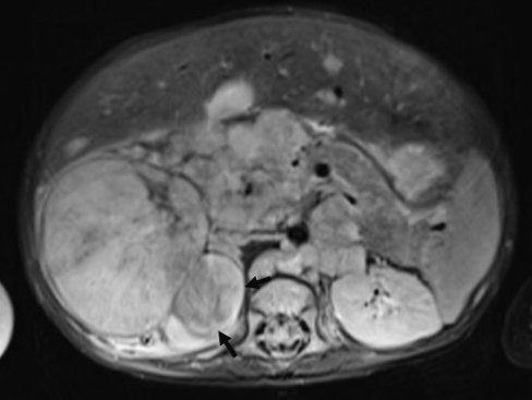
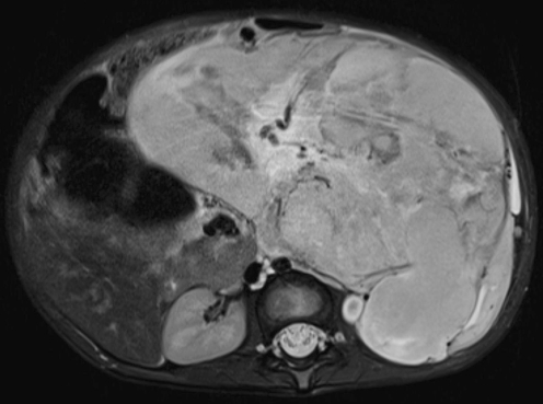
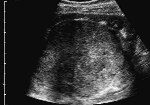
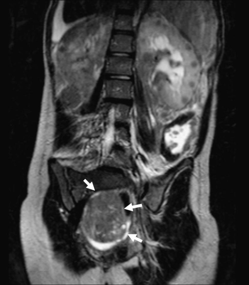
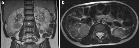
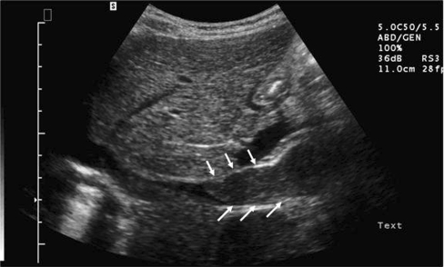
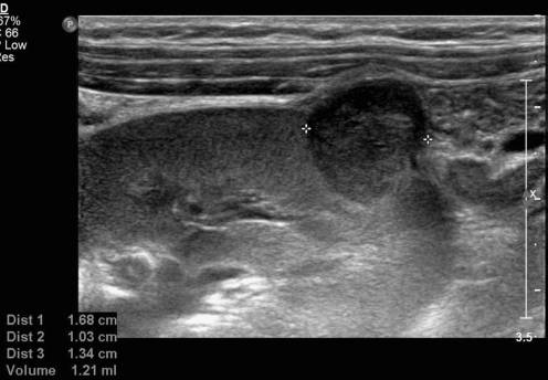
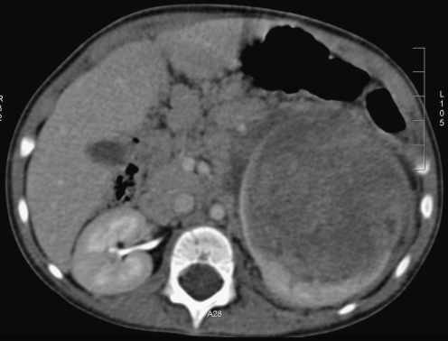
Primitive malignant renal tumours comprise 6% of all childhood cancers. Wilms tumour (WT) or nephroblastoma is the most frequent type accounting for more than 90%. Imaging alone cannot differentiate between these tumours with certainty but it plays an important role in screening, diagnostic workup, assessment of therapy response, preoperative evaluation and follow-up. The outcome of WT after therapy is excellent with an overall survival around 90%. In tumours such as those where the outcome is extremely good, focus can be shifted to a risk-based stratification to maintain excellent outcome in children with low risk tumours while improving quality of life and decreasing toxicity and costs. This review will discuss the imaging issues for WT from the European perspective and briefly discuss the characteristics of other malignant renal tumours occurring in children and new imaging techniques with potential in this matter.
Figures


Similar articles
-
Diagnostic utility of Wilms' tumour-1 protein (WT-1) immunostaining in paediatric renal tumours.Indian J Med Res. 2016 May;143(Supplement):S59-S67. doi: 10.4103/0971-5916.191776. Indian J Med Res. 2016. PMID: 27748279 Free PMC article.
-
Imaging of renal tumours in infancy and childhood.Eur Radiol. 2003 Dec;13 Suppl 4:L116-29. doi: 10.1007/s00330-003-2001-x. Eur Radiol. 2003. PMID: 15018176 Review.
-
Renal and adrenal tumours in children.Cancer Imaging. 2007 Mar 5;7(1):41-51. doi: 10.1102/1470-7330.2007.0007. Cancer Imaging. 2007. PMID: 17339140 Free PMC article. Review.
-
Incidence, treatment, and outcomes of primary and recurrent Non-Wilms renal tumors in children: Report of 109 patients treated at a single institution.J Pediatr Urol. 2020 Aug;16(4):475.e1-475.e9. doi: 10.1016/j.jpurol.2020.05.168. Epub 2020 Jun 5. J Pediatr Urol. 2020. PMID: 32620510
-
[Nephroblastoma in children aged less than 6 months at diagnosis].Med Wieku Rozwoj. 2003 Jul-Sep;7(3):347-57. Med Wieku Rozwoj. 2003. PMID: 14963342 Polish.
Cited by
-
CT and MRI appearances and radiologic staging of pediatric renal cell carcinoma.Pediatr Radiol. 2012 Apr;42(4):410-7; quiz 513-4. doi: 10.1007/s00247-011-2319-5. Epub 2012 Jan 17. Pediatr Radiol. 2012. PMID: 22249598
-
A comparison between ultrasonography and single-phase computed tomography for preoperative assessment of solid abdominal tumors in children.J Pediatr (Rio J). 2023 Jan-Feb;99(1):17-22. doi: 10.1016/j.jped.2022.05.003. Epub 2022 Jun 17. J Pediatr (Rio J). 2023. PMID: 35718001 Free PMC article.
-
Position paper: Rationale for the treatment of children with CCSK in the UMBRELLA SIOP-RTSG 2016 protocol.Nat Rev Urol. 2018 May;15(5):309-319. doi: 10.1038/nrurol.2018.14. Epub 2018 Feb 27. Nat Rev Urol. 2018. PMID: 29485128 Review.
-
Diagnostic value of pseudocapsule ring hyperenhancement in contrast-enhanced ultrasound in renal focal lesions.Ir J Med Sci. 2023 Aug;192(4):1631-1636. doi: 10.1007/s11845-023-03320-x. Epub 2023 Mar 12. Ir J Med Sci. 2023. PMID: 36907918
-
Flank pain and gross hematuria: an uncommon presentation of a rare disease: Answers.Pediatr Nephrol. 2021 Jul;36(7):2049-2051. doi: 10.1007/s00467-020-04900-0. Epub 2021 Jan 13. Pediatr Nephrol. 2021. PMID: 33439352 No abstract available.
References
-
- SIOP (2001) Nephroblastoma clinical trial and study protocol. In: Oncology ISoP (ed), pp 1–170
Publication types
MeSH terms
LinkOut - more resources
Full Text Sources
Medical