. 2010 May;51(5):1810-9.
doi: 10.1002/hep.23626.

Generation of endoderm-derived human induced pluripotent stem cells from primary hepatocytes

Affiliations

Generation of endoderm-derived human induced pluripotent stem cells from primary hepatocytes

Hua Liu et al. Hepatology. 2010 May.

Abstract

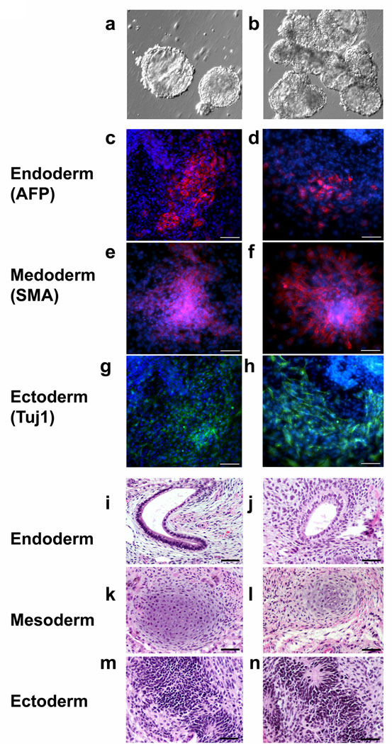
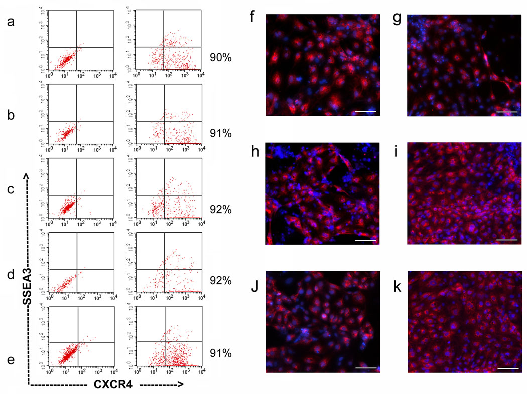
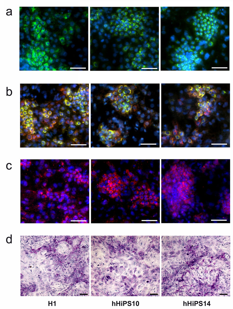
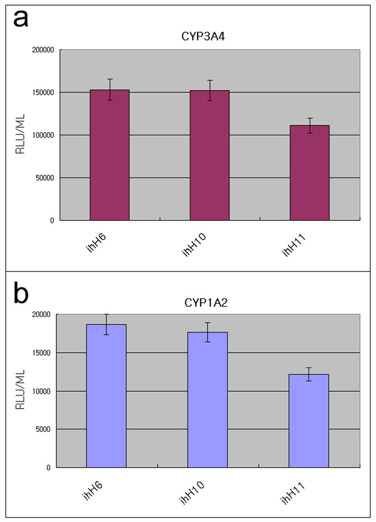
Recent advances in induced pluripotent stem (iPS) cell research have significantly changed our perspective on regenerative medicine. Patient-specific iPS cells have been derived not only for disease modeling but also as sources for cell replacement therapy. However, there have been insufficient data to prove that iPS cells are functionally equivalent to human embryonic stem (hES) cells or are safer than hES cells. There are several important issues that need to be addressed, and foremost are the safety and efficacy of human iPS cells of different origins. Human iPS cells have been derived mostly from cells originating from mesoderm and in a few cases from ectoderm. So far, there has been no report of endoderm-derived human iPS cells, and this has prevented comprehensive comparative investigations of the quality of human iPS cells of different origins. Here we show for the first time reprogramming of human endoderm-derived cells (i.e., primary hepatocytes) to pluripotency. Hepatocyte-derived iPS cells appear indistinguishable from hES cells with respect to colony morphology, growth properties, expression of pluripotency-associated transcription factors and surface markers, and differentiation potential in embryoid body formation and teratoma assays. In addition, these cells are able to directly differentiate into definitive endoderm, hepatic progenitors, and mature hepatocytes.

Conclusion: The technology to develop endoderm-derived human iPS cell lines, together with other established cell lines, will provide a foundation for elucidating the mechanisms of cellular reprogramming and for studying the safety and efficacy of differentially originated human iPS cells for cell therapy. For the study of liver disease pathogenesis, this technology also provides a potentially more amenable system for generating liver disease-specific iPS cells.


Figures

Figure 1
Figure 1. Human hepatocyte derived iPS (hHiPS) cell colony formation and characterization
(a) Phase contrast image and (b) albumin expression of primary human hepatocytes before iPS reprogramming. ×200, Scale bars: 50 µm. (c) A diagram of hHiPS generation protocol. (d) Typical example of a small ES cell–like TRA-1-60 (red) positive colony adjacent to a TRA-1-60 negative non-iPS colony (arrow). (e) Typical ES cell–like TRA-1-60 positive iPS cell colony before harvest. ×100, Scale bars: 100 µm. (f) Representative immunofluorescence analysis of one hHiPS cell line (hHiPS10) growing on Matrigel. Clear expression of the ES cell surface antigens SSEA4 and TRA-1-60, the nuclear transcription factors OCT4 and NANOG are observed (×200). Scale bars: 50 µm.
Figure 2
Figure 2. hHiPS cells can differentiate into all three primary germ layers in vitro and in vivo
(a, b) Embryoid bodies derived from hHiPS 10, and 11 lines, respectively. (c–h) In vitro differentiation of hHiPS cells into all three primary germ cell layers. After generation of embryoid bodies hHiPS cells spontaneously differentiated into endoderm (c, d) (α-fetoprotein-positive, red), mesoderm (e, f) (smooth muscle actin-positive, red) and ectoderm (g, h) (TuJ1-positive neuronal cells, green). c, e, g: hHiPS10 and d, f, h: hHiPS11. Blue nuclear staining is DAPI. ×100, Scale bars, 100 µm. (i–n) Spontaneous differentiation into all three germ layers is evident in teratomas. (i, j) endoderm, (k, l) mesoderm, (m, n) ectoderm derived from hHiPS10 and hHiPS14. ×200, Scale bars, 50 µm.
Figure 3
Figure 3. Differentiation of hHiPS cells into DE and hepatic progenitors
Efficient endoderm induction of human iPS cells and ES cells in the presence of 100 ng/ml Activin A. The FACS analysis showed that at least 90% of the induced cells expressed the definitive endoderm marker CXCR4. H9 (a), H1 (b), and hHiPS6, 10, 11 (c, d, e) at 5 days post-initial Activin A treatment. Immunofluorescence analysis of the expression of AFP in differentiating cells at 10 days post-initial Activin A treatment (f-k, H1, H9, hHiPS6, 10, 11, and 14, respectively). ×100. Scale bars, 100 µm.
Figure 4
Figure 4. Differentiation of hHiPS cells into mature hepatocytes
The differentiated cells possessed proteins indicative of functional hepatocytes, including ALB, green (a), ALB (green) + AAT (red) (b), and CYP3A4 (pink) (c) at day 20 after differentiation initiation. ×200, Scale bars, 100 µm. (d) Glycogen storage at day 20. Periodic acid-Schiff assay was performed on differentiating cells at 20 days post-initial Activin A treatment. Nuclei were counterstained with hematoxylin (blue). Glycogen storage is indicated by pink or dark red cytoplasm. ×100, Scale bar = 100 µm.
Figure 5
Figure 5. CYP450 metabolism in hHiPS cell derived mature hepatocytes
hHiPS cell derived mature hepatocytes display cytochrome P450 metabolism. iPS cell-derived hepatocytes were incubated with hepatocyte culture medium supplemented with CYP3A4 or CYP1A2 pGlo substrates (Promega) as per manufacturer’s instructions. At 4 hours after treatment, 50 uL of culture medium was removed and read on a luminometer (GLOMAX). CYP1A2 and CYP3A4 activity is expressed as relative light units (RLU)/ml of culture medium (n=6). All three hHiPS cell lines (ihH6, 10, 11) exhibited both CYP450 enzyme activities.

Comment in

References

    1. Chin MH, Mason MJ, Xie W, Volinia S, Singer M, Peterson C, et al. Induced pluripotent stem cells and embryonic stem cells are distinguished by gene expression signatures. Cell Stem Cell. 2009;5:111–123. - PMC - PubMed
    1. Marchetto MCN, Yeo GW, Kainohana O, Marsala M, Gage FH, Muotri AR. Transcriptional Signature and Memory Retention of Human-Induced Pluripotent Stem Cells. PLoS ONE. 2009;4:e7076. - PMC - PubMed
    1. Miura K, Okada Y, Aoi T, Okada A, Takahashi K, Okita K, et al. Variation in the safety of induced pluripotent stem cell lines. Nat Biotechnol. 2009;27:743–745. - PubMed
    1. Deng J, Shoemaker R, Xie B, Gore A, LeProust EM, Antosiewicz-Bourget J, et al. Targeted bisulfite sequencing reveals changes in DNA methylation associated with nuclear reprogramming. Nat Biotechnol. 2009;27:353–360. - PMC - PubMed
    1. Doi A, Park IH, Wen B, Murakami P, Aryee MJ, Irizarry R, et al. Differential methylation of tissue- and cancer-specific CpG island shores distinguishes human iPSCs, embryonic stem cells and fibroblasts. Nat Genet. 2009;41:1350–1353. - PMC - PubMed

Publication types

MeSH terms