

In vivo near-IR imaging of approximal dental decay at 1,310 nm
- PMID: 20432277
- PMCID: PMC3195334
- DOI: 10.1002/lsm.20913
In vivo near-IR imaging of approximal dental decay at 1,310 nm
Abstract
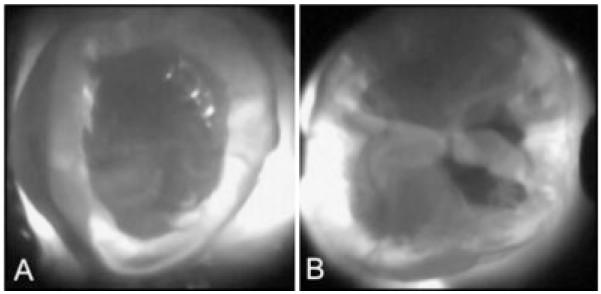
Objective: The high transparency of dental enamel in the near-IR (NIR) light at 1,310-nm can be exploited for imaging dental caries without the use of ionizing radiation (X-rays). We present the results of the first in vivo imaging study in which NIR images were acquired of approximal contact surfaces.
Methods: NIR imaging hand-pieces were developed and attached to a compact InGaAs focal plane array and subsequently used to acquire in vivo NIR images of 33 caries lesions on 18 test subjects. The carious lesions were discernible on bitewing radiographs, but were not visible upon clinical examination.
Results: NIR images were acquired in vivo from three directions and the majority of lesions examined were too small to require restoration, based on accepted bitewing radiograph criteria. All but one of the 33 lesions examined were successfully imaged from at least one direction.
Conclusion: This first in vivo study of imaging at the 1,310-nm wavelength region shows that NIR imaging has great potential as a screening tool for the detection of approximal lesions without the use of ionizing radiation.
(c) 2010 Wiley-Liss, Inc.
Figures


References
-
- Jones RS, Huynh GD, Jones GC, Fried D. Near-IR trans-illumination at 1310-nm for the imaging of early dental caries. Opt Expr. 2003;11(18):2259–2265. - PubMed
-
- Bühler CM, Ngaotheppitak P, Fried D. Imaging of occlusal dental caries (decay) with near-IR light at 1310-nm. Opt Expr. 2005;13(2):573–582. - PubMed
-
- Fried D, Featherstone JDB, Glena RE, Seka W. The nature of light scattering in dental enamel and dentin at visible and near-IR wavelengths. Appl Opt. 1995;34(7):1278–1285. - PubMed
-
- Jones RS, Fried D. Lasers in dentistry VIII. SPIE; San Jose: 2002. Attenuation of 1310-nm and 1550-nm laser light through sound dental enamel; pp. 187–190.
-
- Darling CL, Huynh GD, Fried D. Light scattering properties of natural and artificially demineralized dental enamel at 1310-nm. J Biomed Opt. 2006;11(3):034023, 034021–034011. - PubMed
MeSH terms
Grants and funding
LinkOut - more resources
Full Text Sources
Other Literature Sources
Medical
Miscellaneous
