. 2010 May;63(5):1383-90.
doi: 10.1002/mrm.22313.

Magnetic nanoparticles for imaging dendritic cells

Affiliations

Magnetic nanoparticles for imaging dendritic cells

Saho Kobukai et al. Magn Reson Med. 2010 May.

Abstract

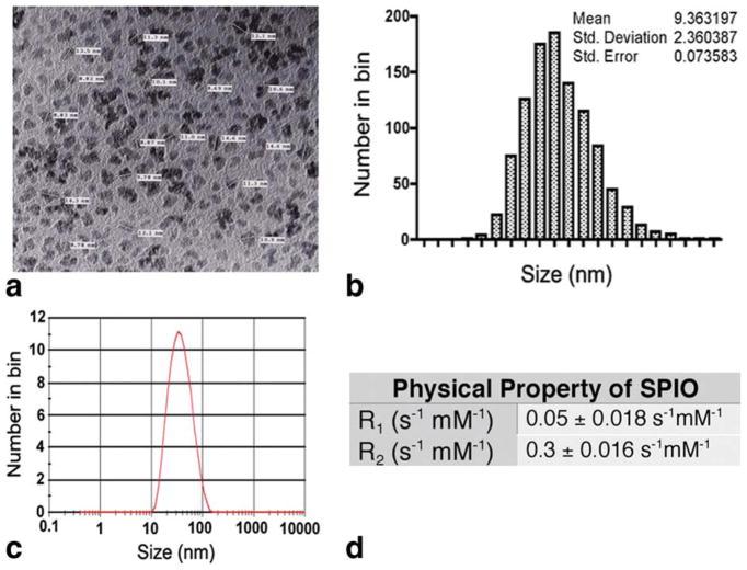
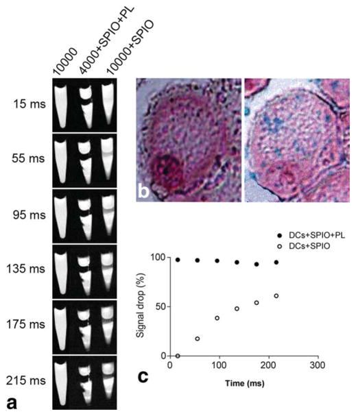
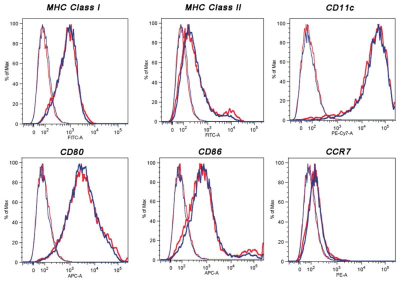
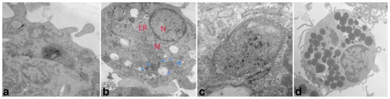
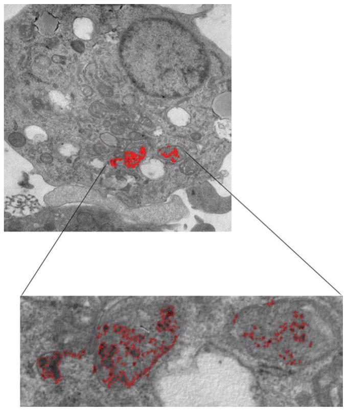
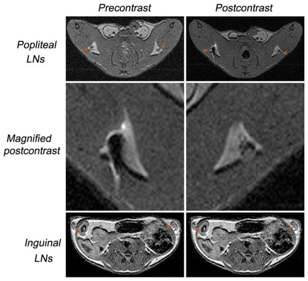
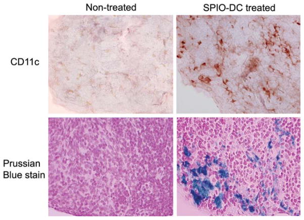
We report the development of superparamagnetic iron oxide (SPIOs) nanoparticles and investigate the migration of SPIO-labeled dendritic cells (DCs) in a syngeneic mouse model using magnetic resonance (MR) imaging. The size of the dextran-coated SPIO is roughly 30 nm, and the DCs are capable of independent uptake of these particles, although not at levels comparable to particle uptake in the presence of a transfecting reagent. On average, with the assistance of polylysine, the particles were efficiently delivered inside DCs within one hour of incubation. The SPIO particles occupy approximately 0.35% of cell surface and are equivalent to 34.6 pg of iron per cell. In vivo imaging demonstrated that the labeled cells migrated from the injection site in the footpad to the corresponding popliteal lymph node. The homing of labeled cells in the lymph nodes resulted in a signal drop of up to 79%. Furthermore, labeling DCs with SPIO particles did not compromise cell function, we demonstrated that SPIO-enhanced MR imaging can be used to track the migration of DCs effectively in vivo.


Figures

FIG. 1
FIG. 1
Physical properties of the synthesized dextran-coated SPIO particles. a: TEM image of the SPIO nanoparticles. b: The distribution of the iron core calculated from the TEM image. c: The overall size of dextran-coated SPIO particles was measured by a Zetasizer. d: The R values.
FIG. 2
FIG. 2
a: Magnetic resonance imaging of gelatin-embedded DCs at 4.7 T after incubation with SPIO for 1 h, with or without PL. The density of cells is represented by the numbers. b: Visualization using Prussian blue staining of the internalized SPIO particles with (right) and without (left) the presence of PL. c: Quantification of the signal drop caused by the presence of SPIO in the gelatin phantom tube as a percentage compared to the control.
FIG. 3
FIG. 3
Cell surface phenotype as determined by flow cytometry of unlabeled (blue) and SPIO-labeled (red) murine-derived DCs. After 1 h of incubation with SPIO in the presence of PL, cells were stained with phycoerythrin-conjugated monoclonal antibodies for CD11c, presentation receptors MHC-I and MHC-II, costimulatory receptors CD80 and CD86, and chemokine receptors CCR7. The left histograms represent unstained labeled and unlabeled cells as controls. The shifted histograms represent stained labeled and unlabeled DCs. The y-axis of each histogram shows the relative cell number; the x-axis shows the log fluorescence intensity.
FIG. 4
FIG. 4
Morphologic analysis of labeled DCs. Cells were incubated with SPIO for 1 h in the presence of PL and then processed for TEM analysis. a: Initial point of entry after membrane association; (b) SPIO-containing vesicles in the cytoplasm (arrowheads); (c) enlarged vesicle in b (star), and (d) control DCs without SPIO.
FIG. 5
FIG. 5
Surface area occupancy of SPIO in a DC was calculated using a program written in house with MATLAB. SPIO vesicles occupy ~0.35% of cell space.
FIG. 6
FIG. 6
Transaxial in vivo multispin-echo MRIs (TE = 10 ms, TR = 2–3 sec) of mice before and after distribution of the labeled cells via the foot pad. The LNs are indicated by arrows. The signal intensity of the popliteal LNs appears darker, suggesting the migration and homing of the DCs. No signal intensity change was detected before or after injection of SPIO-labeled DCs in inguinal LNs.
FIG. 7
FIG. 7
Immunohistochemical and iron staining (both ×40) of consecutive tissue slides of the popliteal LNs, either untreated or treated, with foot pad injection of SPIO-DCs over the course of 24 h.

References

    1. Larsson M, Majeed M, Ernst JD, Magnusson KE, Stendahl O, Forsum U. Role of annexins in endocytosis of antigens in immature human dendritic cells. Immunology. 1997;92:501–511. - PMC - PubMed
    1. Mohty M, Olive D, Gaugler B. Leukemic dendritic cells: potential for therapy and insights towards immune escape by leukemic blasts. Leukemia. 2002;16:2197–2204. - PubMed
    1. Ni K, O’Neill HC. The role of dendritic cells in T cell activation. Immunol Cell Biol. 1997;75:223–230. - PubMed
    1. Fernandez NC, Lozier A, Flament C, Ricciardi-Castagnoli P, Bellet D, Suter M, Perricaudet M, Tursz T, Maraskovsky E, Zitvogel L. Dendritic cells directly trigger NK cell functions: cross-talk relevant in innate anti-tumor immune responses in vivo. Nat Med. 1999;5:405–411. - PubMed
    1. Yang L, Carbone DP. Tumor-host immune interactions and dendritic cell dysfunction. Adv Cancer Res. 2004;92:13–27. - PubMed

Publication types