. 2010 Sep;224(3):649-57.
doi: 10.1002/jcp.22163.

Mechanistic and signaling analysis of Muc4-ErbB2 signaling module: new insights into the mechanism of ligand-independent ErbB2 activity

Affiliations

Mechanistic and signaling analysis of Muc4-ErbB2 signaling module: new insights into the mechanism of ligand-independent ErbB2 activity

Goldi A Kozloski et al. J Cell Physiol. 2010 Sep.

Abstract

The membrane mucin Muc4 is aberrantly expressed in numerous epithelial carcinomas and is currently used as a cancer diagnostic and prognostic tool. Muc4 can also potentiate signal transduction by modulating differential ErbB2 phosphorylation in the absence and in the presence of the ErbB3 soluble ligand heregulin (HRG-beta1). These features of Muc4 suggest that Muc4 is not merely a cancer marker, but an oncogenic factor with a unique-binding/activation relationship with the receptor ErbB2. In the present study, we examined the signaling mechanisms that are associated with the Muc4-ErbB2 module by analyzing ErbB2 differential signaling in response to Muc4 expression. Our study was carried out in the A375 human melanoma and BT-474 breast cancer cell lines as our model systems. Quantitative and comparative signaling modulations were evaluated by immunoblot using phospho-specific antibodies, and densitometry analysis. Signaling complex components were identified by chemical cross-linking, fractionation by gel filtration, immunoprecipitation, and immunoblotting. Activated downstream signaling pathways were analyzed by an antibody microarray screen and immunoblot analyses. Our results indicate that Muc4 modulates ErbB2 signaling potential significantly by stabilizing and directly interacting with the ErbB2-ErbB3 heterodimer. Further analyses indicate that Muc4 promotes ErbB2 autocatalysis, but it has no effect on ErbB3 phosphorylation, although the chemical cross-linking data indicated that the signaling module is composed of Muc4, ErbB2, and ErbB3. Our microarray analysis indicates that Muc4 expression promotes cell migration by increasing the phosphorylation of the focal adhesion kinase and also through an increase in the levels of beta-catenin.


Figures

FIGURE 1
FIGURE 1
Muc4 modulates ErbB2 signaling potential . A. Muc4 forms a stable complex with phosphorylated ErbB2. Proteins from RIPA extracts of A375 Muc4-transfected cells with or without Muc4 (48 h) were subjected to immunoprecipitation with anti-rCpep, a Muc4 pAb targeting the cytoplasmic portion of Muc4, or with the pre-immune serum (−). Immunoprecipitates were immunoblotted with anti-Muc4, anti pY1248-ErbB2, and anti-ErbB3 antibodies. The control immunoprecipitation lane (+) in the ErbB3 immunoblot was carried out with the anti-ErbB3 (C-17) polyclonal antibody. The input control is an immunoblot of the lysates with anti-Muc4 and anti β-actin antibodies. B. Muc4 expression augments ErbB2 phosphorylation signal magnitude significantly (p=0.0002) without changing ErbB2 receptor levels. A375 Muc4-transfected (Rep3 clone) cells with or without Muc4 (48 h) and starved (0.1% FBS) for 24 h were immunoblotted in triplicates with anti-Muc4, anti ErbB2-Y1248, anti-ErbB2, and anti β-actin antibodies. Quantitative analysis of the ErbB2-Y1248 signal intensity is also shown. The signal intensity was calculated using the ImageJ NIH densitometry software. The ErbB2-Y1248 signal was measured, normalized with the β-actin signal, and expressed as the mean ± standard deviation for a series of at least 3 experiments. Student’s t tests were used to compare mean values as appropriate. P values < 0.05 were considered to represent significant differences. C. Muc4 expression stabilizes ErbB2 phosphorylation signal under heregulin ligand (HRG-1β) treatment. A375 Muc4-transfected (Rep 3 clone) cells with or without Muc4 (48 h) and starved (0.1% FBS) for 24 h, were incubated at 37°C with HRG-1β [50 nM] for the indicated times. Quantitative analysis of ErbB2-Y1248 immunoblot band density is shown. Using densitometry software (ImageJ NIH), the signal was measured, normalized with β-actin signal, and then expressed as the mean ± standard deviation for a series of at least three experiments. Circles represent Muc4-Off cells treated with ligand, and squares, Muc4-On cells treated with ligand.
FIGURE 1
FIGURE 1
Muc4 modulates ErbB2 signaling potential . A. Muc4 forms a stable complex with phosphorylated ErbB2. Proteins from RIPA extracts of A375 Muc4-transfected cells with or without Muc4 (48 h) were subjected to immunoprecipitation with anti-rCpep, a Muc4 pAb targeting the cytoplasmic portion of Muc4, or with the pre-immune serum (−). Immunoprecipitates were immunoblotted with anti-Muc4, anti pY1248-ErbB2, and anti-ErbB3 antibodies. The control immunoprecipitation lane (+) in the ErbB3 immunoblot was carried out with the anti-ErbB3 (C-17) polyclonal antibody. The input control is an immunoblot of the lysates with anti-Muc4 and anti β-actin antibodies. B. Muc4 expression augments ErbB2 phosphorylation signal magnitude significantly (p=0.0002) without changing ErbB2 receptor levels. A375 Muc4-transfected (Rep3 clone) cells with or without Muc4 (48 h) and starved (0.1% FBS) for 24 h were immunoblotted in triplicates with anti-Muc4, anti ErbB2-Y1248, anti-ErbB2, and anti β-actin antibodies. Quantitative analysis of the ErbB2-Y1248 signal intensity is also shown. The signal intensity was calculated using the ImageJ NIH densitometry software. The ErbB2-Y1248 signal was measured, normalized with the β-actin signal, and expressed as the mean ± standard deviation for a series of at least 3 experiments. Student’s t tests were used to compare mean values as appropriate. P values < 0.05 were considered to represent significant differences. C. Muc4 expression stabilizes ErbB2 phosphorylation signal under heregulin ligand (HRG-1β) treatment. A375 Muc4-transfected (Rep 3 clone) cells with or without Muc4 (48 h) and starved (0.1% FBS) for 24 h, were incubated at 37°C with HRG-1β [50 nM] for the indicated times. Quantitative analysis of ErbB2-Y1248 immunoblot band density is shown. Using densitometry software (ImageJ NIH), the signal was measured, normalized with β-actin signal, and then expressed as the mean ± standard deviation for a series of at least three experiments. Circles represent Muc4-Off cells treated with ligand, and squares, Muc4-On cells treated with ligand.
FIGURE 2
FIGURE 2
Muc4 modulates ErbB2 signaling potential by stabilizing and directly interacting with the ErbB2-ErbB3 heterodimer. A. Muc4 promotes stable ErbB2-ErbB3 interaction. Proteins from RIPA extracts of A375 cells with or without Muc4 (48 h) were subjected to immunoprecipitation with anti-ErbB3 (C-17) antibody. Immunoprecipitates were immunoblotted with anti-ErbB3, anti-Muc4, and anti-ErbB2 antibodies. The input control is an immunoblot of the lysates with anti-Muc4 and anti β-actin antibodies. B. Muc4 expression results in Muc4-ErbB2-ErbB3 complexes. Cellular chemical cross-linking immunoblots. A375 Muc4-transfected (Rep 3 clone) cells with or without Muc4 (48 h) were crossed linked with chemical cross-linking agents. i). With BS3 [1 mM] for 2 h on ice, and quenched with Tris-HCl [20–50 mM], pH 7.5 at room temperature for 15 min. Yellow arrows indicate cross-linked products. Cell lysates were separated on 4–15% gradient gels, and blotted with anti-Muc4, anti-ErbB2, anti-ErbB3, and β-actin antibodies. ii). With the reversible chemical cross linking analogue, DTSSP, at [1 mM] for 2 h, quenched with Tris-HCl [20–50 mM], pH 7.5 at room temperature for 15 min. Cross-linked cells were lysed in RIPA buffer without phosphatase inhibitors or dithiothreitol. Cleared lystes were loaded by FPLC (1 mL/min) onto a high resolution Superose 6 analytical gel filtration column equilibrated at 4°C in RIPA buffer without Triton X-100, inhibitors or dithiothreitol. Collected fractions were TCA-precipitated and separated on 4–15% gradient gels. Muc4-Off fractions were treated with 5% β-mercaptoethanol-containing SDS-PAGE sample buffer (top). Muc4-On fractions (bottom) were loaded side by side in duplicates with native SDS-PAGE sample buffer to represent non-cleaved (NC) samples, or with 5% β-mercaptoethanol containing SDS-PAGE sample buffer to examine the cleaved complex components (C). Samples were then immunoblotted with the indicated antibodies.
FIGURE 2
FIGURE 2
Muc4 modulates ErbB2 signaling potential by stabilizing and directly interacting with the ErbB2-ErbB3 heterodimer. A. Muc4 promotes stable ErbB2-ErbB3 interaction. Proteins from RIPA extracts of A375 cells with or without Muc4 (48 h) were subjected to immunoprecipitation with anti-ErbB3 (C-17) antibody. Immunoprecipitates were immunoblotted with anti-ErbB3, anti-Muc4, and anti-ErbB2 antibodies. The input control is an immunoblot of the lysates with anti-Muc4 and anti β-actin antibodies. B. Muc4 expression results in Muc4-ErbB2-ErbB3 complexes. Cellular chemical cross-linking immunoblots. A375 Muc4-transfected (Rep 3 clone) cells with or without Muc4 (48 h) were crossed linked with chemical cross-linking agents. i). With BS3 [1 mM] for 2 h on ice, and quenched with Tris-HCl [20–50 mM], pH 7.5 at room temperature for 15 min. Yellow arrows indicate cross-linked products. Cell lysates were separated on 4–15% gradient gels, and blotted with anti-Muc4, anti-ErbB2, anti-ErbB3, and β-actin antibodies. ii). With the reversible chemical cross linking analogue, DTSSP, at [1 mM] for 2 h, quenched with Tris-HCl [20–50 mM], pH 7.5 at room temperature for 15 min. Cross-linked cells were lysed in RIPA buffer without phosphatase inhibitors or dithiothreitol. Cleared lystes were loaded by FPLC (1 mL/min) onto a high resolution Superose 6 analytical gel filtration column equilibrated at 4°C in RIPA buffer without Triton X-100, inhibitors or dithiothreitol. Collected fractions were TCA-precipitated and separated on 4–15% gradient gels. Muc4-Off fractions were treated with 5% β-mercaptoethanol-containing SDS-PAGE sample buffer (top). Muc4-On fractions (bottom) were loaded side by side in duplicates with native SDS-PAGE sample buffer to represent non-cleaved (NC) samples, or with 5% β-mercaptoethanol containing SDS-PAGE sample buffer to examine the cleaved complex components (C). Samples were then immunoblotted with the indicated antibodies.
FIGURE 3
FIGURE 3
Muc4 has no effect on ErbB3 phosphorylation. A375 Muc4-transfected (Rep 3 clone) cells with or without Muc4 (48 h) and starved (0.1% FBS) for 24 h were incubated at 37°C with HRG-1β [50 nM] for 3 min. Cleared lysates were loaded in triplicates and immunoblotted with anti-ErbB3, anti-ErbB Y1289, and anti β-actin antibodies.
FIGURE 4
FIGURE 4
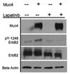
Muc4 promotes ErbB2 autocatalysis. BT-474 cells (Muc4-0ff) and BT-474 cells transfected with Muc4-Rep 3 plasmid for 48 h (Muc4-On) were serum starved (24 h) and incubated with the kinase inhibitor, Lapatinib-GW572016 [0.2 µM] for 6 h. Cells were rinsed twice with ice-cold PBS saline and lysed in RIPA buffer pH 7.2. Cleared lysates were immunoblotted with anti-Muc4, anti ErbB2-Y1248, anti-ErbB2, and anti β-actin antibodies.
FIGURE 5
FIGURE 5
Muc4 expression promotes cell migration signaling by increasing the phosphorylation of focal adhesion kinase (FAK). Immunoblot validation analyses of the microarry screen results. Proteins from RIPA extracts of A375 Muc4-transfected cells induced or not to express Muc4 (48 h) were immunoblotted with the indicated signaling antibodies. Normalized signal units were calculated using the ImageJ NIH densitometry software, and normalized with the corresponding β-actin singal.

Similar articles

Cited by

References

    1. Alvarado D, Klein DE, Lemmon MA. Nature. 2009;461(7261):287–291. - PMC - PubMed
    1. Burgess AW. Growth Factors. 2008;26(5):263–274. - PubMed
    1. Carraway KL, III, Rossi EA, Komatsu M, Price-Schiavi SA, Huang D, Guy PM, Carvajal ME, Fregien N, Carraway CAC, Carraway KL. J. Biol. Chem. 1999;274(9):5263–5266. - PubMed
    1. Carraway KL, Theodoropoulos G, Kozloski GA, Carothers Carraway CA. Future Oncol. 2009;5(10):1631–1640. - PMC - PubMed
    1. Chaturvedi P, Singh AP, Chakraborty S, Chauhan SC, Bafna S, Meza JL, Singh PK, Hollingsworth MA, Mehta PP, Batra SK. Cancer Res. 2008;68(7):2065–2070. - PMC - PubMed

Publication types