. 2010 Jun 1;49(21):4516-23.
doi: 10.1021/bi1003684.

Control of erythrocyte membrane-skeletal cohesion by the spectrin-membrane linkage

Affiliations

Control of erythrocyte membrane-skeletal cohesion by the spectrin-membrane linkage

Lionel Blanc et al. Biochemistry. .

Abstract

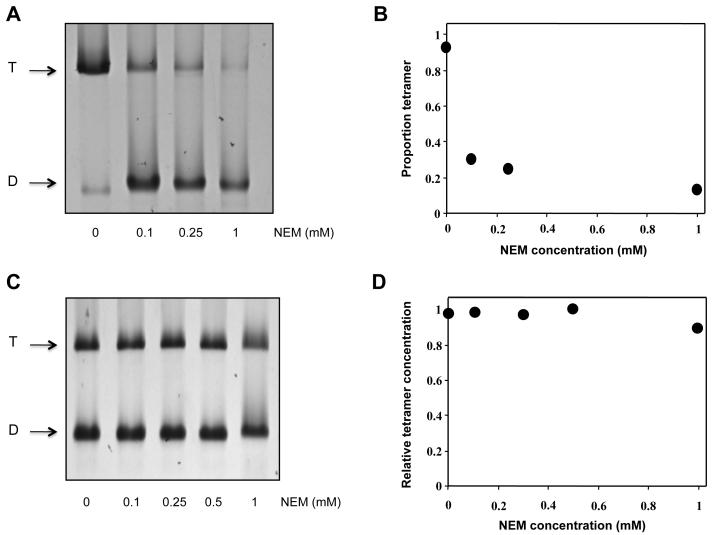
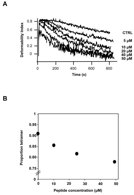
Spectrin tetramer is the major structural member of the membrane-associated skeletal network of red cells. We show here that disruption of the spectrin-ankyrin-band 3 link to the membrane leads to dissociation of a large proportion of the tetramers into dimers. Noncovalent perturbation of the linkage was induced by a peptide containing the ankyrin-binding site of the spectrin beta-chain, and covalent perturbation by treatment with the thiol reagent, N-ethylmaleimide (NEM). This reagent left the intrinsic self-association capacity of the spectrin dimers unaffected and disturbed only the ankyrin-band 3 interaction. The dissociation of spectrin tetramers on the membrane into functional dimers was confirmed by the binding of a spectrin peptide directed against the self-association sites. Dissociation of the tetramers resulted, we infer, from detachment of the proximal ends of the constituent dimers from the membrane, thereby reducing their proximity to one another and thus weakening their association. The measured affinity of the interaction of the peptides with the free dimer ends on the membrane permits an estimate of the equilibrium between intact and dissociated tetramers on the native membrane. This indicates that in the physiological state the equilibrium proportion of the dissociated tetramers may be as high as 5-10%. These findings enabled us to identify an additional important functional role for the spectrin-ankyrin-band 3 link in regulating spectrin self-association in the red cell membrane.


Figures

Fig 1
Fig 1. Effect of N-ethylmaleimide on the association state of spectrin in the red cell membrane and in solution
A. Native polyacrylamide gel showing separation of spectrin dimer (D) and tetramer (T), after extraction from cells treated with the indicated concentrations of NEM. B. Proportion of spectrin converted to dimer in situ, determined by densitometry of the gels. C. Polyacrylamide gel and D. densitometric evaluation of equilibrium mixture of spectrin dimer and tetramer after treatment in solution with the indicated concentrations of NEM.
Fig 2
Fig 2. Equilibria and kinetics of binding of peptides to the self-association sites of spectrin in the red cell membrane
Kinetics of binding to native (open circles) and NEM-reacted (filled circles) red cell ghosts at 24°C (A.) and at 37°C (B.) of αII spectrin peptide, directed at the spectrin dimer self-association site. C. Equilibrium binding at 37°C of βI spectrin peptide, directed at the dimer self-association sites. The curves are best-fits, giving apparent association constants, Kapp = 1.3 (± 0.1) × 104 and 1.5 (± 0.2) × 103 M−1, respectively, uncorrected for dimer-tetramer equilibrium (see text).
Fig 3
Fig 3. Binding of native spectrin to inside-out membrane vesicles from cells treated with N-ethylmaleimide, and of spectrin from cells thus treated to vesicles of untreated cells
Spectrins bound to vesicles as a function of spectrin concentration: vesicles and spectrin from untreated cells (dashed circles). Spectrin from NEM-treated cells and vesicles from untreated cells (open circles), spectrin from untreated cells and vesicles from NEM-treated cells (filled circles). The curves are best fits for binding to a population of identical and independent sites, giving saturation binding titres of 2.8 + 0.3, 2.6 + 0.2 and 2.4 + 0.3 nM respectively, with corresponding apparent association constants of 0.6 + (0.13) × 106, 1.5 (+ 0.3) × 106, and 1.0 (+ 0.3) × 106 M−1.
Fig 4
Fig 4. Extraction of ankyrin from inside-out vesicles of cells before and after treatment with N-ethylmaleimide as a function of ionic strength of the extracting medium
A. Residual ankyrin on the membranes as a function of ionic strength of the extraction buffer in spectrin-depleted inside-out vesicles before and after NEM treatment as documented by western blot analysis. B. Fractions of ankyrin remaining in the vesicles after extraction: filled circles refer to NEM-treated, and open circles to control cells. Note that in the former, the proportion of residual protein refers to the ankyrin present in the vesicles as prepared, which have already lost nearly half their ankyrin in the course of preparation.
Fig 5
Fig 5. Effect of incorporation of β-spectrin peptide that binds to ankyrin (βIR(14-15) on membrane mechanical stability and spectrin dimer- tetramer equilibrium
A. membrane mechanical stability of the resealed ghosts was measured by ektacytometry. Membrane stability, expressed as the rate of decline in deformability index, DI, diminishes (decay curve displaced toward shorter times) with increasing concentrations of peptide incorporated into cells. B. Densitometric evaluation of native electrophoretic gels of spectrin extracted from cells treated with peptide, showing partial dissociation of spectrin tetramers.
Fig 6
Fig 6. Schematic model showing suggested consequences of disruption of the spectrin ankyrin-band 3 bridge on spectrin self-association
The spectrin α-chains are shown in light green, the β-chains in dark green. Inset: The interactions between ankyrin and β–spectrin are highlighted. Zu5, a subdomain of ankyrin, binds to β–spectrin through the terminal part of R14 and most of the R15, and has been identified as the minimal binding domain for β–spectrin (27, 39).

References

    1. Chasis JA, Mohandas N. Erythrocyte membrane deformability and stability: two distinct membrane properties that are independently regulated by skeletal protein associations. J. Cell. Biol. 1986;103:343–350. - PMC - PubMed
    1. Mohandas N, Gallagher PG. Red cell membrane: past, present, and future. Blood. 2008;112:3939–3948. - PMC - PubMed
    1. Johnson CP, Tang HY, Carag C, Speicher DW, Discher DE. Forced unfolding of proteins within cells. Science. 2007;317:663–666. - PMC - PubMed
    1. Smith DK, Palek J. Sulfhydryl reagents induce altered spectrin self-association, skeletal instability, and increased thermal sensitivity of red cells. Blood. 1983;62:1190–1196. - PubMed
    1. Fischer TM, Haest CW, Stöhr M, Kamp D, Deuticke B. Selective alteration of erythrocyte deformabiliby by SH-reagents: evidence for an involvement of spectrin in membrane shear elasticity. Biochim. Biophys. Acta. 1978;510:270–282. - PubMed

Publication types