Review
. 2010 Oct;6(5):634-41.
doi: 10.1016/j.nano.2010.04.005. Epub 2010 Apr 28.

Viral nanoparticles as platforms for next-generation therapeutics and imaging devices

Affiliations
Review

Viral nanoparticles as platforms for next-generation therapeutics and imaging devices

Nicole F Steinmetz. Nanomedicine. 2010 Oct.

Abstract

Nanomaterials have been developed for potential applications in biomedicine, such as tissue-specific imaging and drug delivery. There are many different platforms under development, each with advantages and disadvantages, but viral nanoparticles (VNPs) are particularly attractive because they are naturally occurring nanomaterials, and as such they are both biocompatible and biodegradable. VNPs can be designed and engineered using both genetic and chemical protocols. The use of VNPs has evolved rapidly since their introduction 20 years ago, encompassing numerous chemistries and modification strategies that allow the functionalization of VNPs with imaging reagents, targeting ligands, and therapeutic molecules. This review discusses recent advances in the design of "smart" targeted VNPs for therapeutic and imaging applications.

From the clinical editor: This review focuses on viral nanoparticles, which are considered attractive naturally occurring nanomaterials due to their inherent biocompatibility and biodegradability. These can be used as imaging reagents, targeting ligands and therapeutic molecules.


Figures

Figure 1
Figure 1. A snapshot of the viral nanoparticles (VNPs) currently being developed for applications in medicine
Icosahedral plant viruses: Brome mosaic virus (BMV), Cowpea chlorotic mottle virus (CCMV), Cowpea mosaic virus (CPMV), Hibiscus chlorotic ringspot virus (HCRSV), Red clover necrotic mottle virus (RCNMV). Icosahedral bacteriophages: MS2 and Qβ, and the filamentous phage M13. Rod-shaped plant viruses: Potato virus X (PVX), Tobacco mosaic virus (TMV). Images of the following VNPs were reproduced from the VIPER database (www.viperdb.scripps.edu): BMV, CCMV, CPMV, RCNMV, MS2, Qβ. The structure of HCRSV was reproduced from Doan DN et al. (2003) J Struct Biol 144(3): 253–261. M13 was reproduced from Khalil AS et al. (2007) PNAS 104(12): 4892–4897. The structure of PVX is from Kendall A et al. (2008) J Virol 82(19): 9546–9554. The cryo-reconstruction of TMV was provided by Bridget Carragher and Clint Potter; data were collected and processed at the National Resource for Automated Molecular Microscopy (NRAMM) at the Scripps Research Institute.
Figure 2
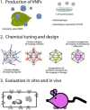
Figure 2. Viral nanotechnology – the assembly line
1. VNPs can be produced in their natural hosts: plants when using plant viruses, bacteria when using bacteriophages, mammalian cells when using mammalian viruses. Heterologous expression of VLPs in bacteria and yeast is also a common production technique. 2. Once purified, chemical tuning and design is carried out to attach and encapsulate molecules that confer different functionalities. 3. The hybrid and functionalized VNP is then evaluated in vitro and in vivo.
Figure 3
Figure 3. Intravital imaging using viral nanoparticles
Fluorescent-labeled CPMV probes (CPMV-A555) in either mouse (A–D) or chick (E) embryos. A+B. Imaging of CPMV-A555 perfused into 11.5-day-old mouse embryo with intact yolk sac (A) and removed yolk sac (B). White boxes indicate the regions magnified in (C) and (D). Comparison of intravital staining intensity over time in the chick embryo (E) using either CPMV-A555 or nanospheres as fluorescent probes. Representative images captured immediately after and 4 h after injection. Reproduced from Lewis JD et al. (2006) Viral nanoparticles as tools for intravital vascular imaging. Nat Med 12: 354–360.
Figure 4
Figure 4. Fluorescent CPMV nanoparticles highlight tumor angiogenesis: intravital imaging in a CAM/HT1080 fibrosarcoma model
A) Bright-field image of HT1080 tumor on-plant on the chick chorioallantoic membrane (CAM) at 7 d. Opaque object is a nylon mesh grid used for quantification of angiogenesis. B) Fluorescence image of tumor on-plant after injection of embryo with CPMV-AF555. C) High magnification image of tumor interior shown in b; tumor microvasculature is clearly observed. D,E) Visualization of HT1080 tumor angiogenesis using CPMV-A555. D) Left, visualization of pre-existing vasculature in the CAM immediately after HT1080 tumor cell injection with CPMV-A555. Middle, GFP-expressing HT1080 tumor bolus under the surface of the CAM. Right, merge. Scale bar, 100 mm. E) Left, visualization of pre-existing CAM vasculature and neovasculature arising from tumor angiogenesis 24 h after tumor-cell injection. Middle, GFP expressing HT1080 tumor bolus. The extensive migration over 24 h indicates a high level of tumor-cell viability. Right, merge. Scale bar, 100 mm. Reproduced from Lewis JD et al. (2006) Viral nanoparticles as tools for intravital vascular imaging. Nat Med 12: 354–360.
Figure 5
Figure 5. VNP-C60 conjugated in cancer cells
(A) HeLa cells only. (B–F) Cells treated with Qβ-PEG-C60-A568 particles. Color key: blue, nuclei (DAPI); red, Qβ-PEG-C60-A568; green, A488-labeled wheat germ agglutinin. (D) Z-section image (1.2 μm deep) recorded along the line shown in (C); step size 0.3 μm. (E, F) Same cell as shown in (D), image reconstructions using Imaris software. Reproduced from Steinmetz NF et al. (2009) Buckyballs meet viral nanoparticles: candidates for biomedicine. JACS 131: 17093–17095.

References

    1. Majoros IJ, Williams CR, Baker JR., Jr. Current dendrimer applications in cancer diagnosis and therapy. Curr Top Med Chem. 2008;8:1165–79. - PubMed
    1. Manchester M, Singh P. Virus-based nanoparticles (VNPs): platform technologies for diagnostic imaging. Adv Drug Deliv Rev. 2006;58:1505–22. - PubMed
    1. Soussan E, Cassel S, Blanzat M, Rico-Lattes I. Drug delivery by soft matter: matrix and vesicular carriers. Angewandte Chemie. 2009;48:274–88. - PubMed
    1. Xing Y, Rao J. Quantum dot bioconjugates for in vitro diagnostics & in vivo imaging. Cancer Biomark. 2008;4:307–19. - PubMed
    1. Portney NG, Ozkan M. Nano-oncology: drug delivery, imaging, and sensing. Anal Bioanal Chem. 2006;384:620–30. - PubMed

Publication types