. 2010 Aug;12(8-9):652-661.
doi: 10.1016/j.micinf.2010.04.008. Epub 2010 Apr 29.

Chlamydial infection of monocytes stimulates IL-1beta secretion through activation of the NLRP3 inflammasome

Affiliations

Chlamydial infection of monocytes stimulates IL-1beta secretion through activation of the NLRP3 inflammasome

Ali A Abdul-Sater et al. Microbes Infect. 2010 Aug.

Abstract

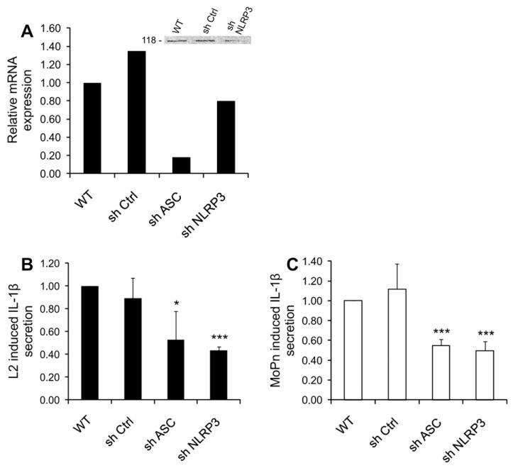
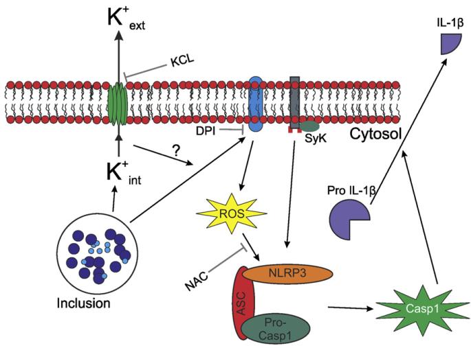
Chlamydia trachomatis infections represent the leading cause of bacterial sexually-transmitted disease in the United States and can cause serious tissue damage leading to infertility and ectopic pregnancies in women. Inflammation and hence the innate immune response to chlamydial infection contributes significantly to tissue damage, particularly by secreting proinflammatory cytokines such as interleukin (IL)-1beta from monocytes, macrophages and dendritic cells. Here we demonstrate that C. trachomatis or Chlamydia muridarum infection of a monocytic cell line leads to caspase-1 activation and IL-1beta secretion through a process requiring the NLRP3 inflammasome. Thus, secretion of IL-1beta decreased significantly when cells were depleted of NLRP3 or treated with the anti-inflammatory inhibitors parthenolide or Bay 11-7082, which inhibit inflammasomes and the transcription factor NF-kappaB. As for other infections causing NRLP3 inflammasome assembly, caspase-1 activation in monocytes is triggered by potassium efflux and reactive oxygen species production. However, anti-oxidants inhibited IL-1beta secretion only partially. Atypically for a bacterial infection, caspase-1 activation during chlamydial infection also involves partially the spleen tyrosine kinase (Syk), which is usually associated with a pathogen recognition receptor for fungal pathogens. Secretion of IL-1beta during infection by many bacteria requires both microbial products from the pathogen and an exogenous danger signal, but chlamydial infection provides both the pathogen-associated molecular patterns and danger signals necessary for IL-1beta synthesis and its secretion from human monocytes. Use of inhibitors that target the inflammasome in animals should therefore dampen inflammation during chlamydial infection.


Figures

Fig. 1
Fig. 1
Chlamydia induces caspase-1 activation in monocytes. THP-1 cells treated with non-target control (sh Control) were infected with C. trachomatis (L2) at an m.o.i. of 5 or C. muridarum (MoPn) at an m.o.i. of 1 for 24 h. (A) Western blot analysis of the lysates was performed to monitor caspase-1 (Casp1) activation using an antibody that detects active caspase-1 (p20). (B) Caspase-1 ELISA was used to quantitatively measure caspase-1 activity in the supernatant of cultured cells. (C) IL-1β ELISA was used to measure IL-1β secretion in supernatants of cultured cells. Error bars represent standard deviation of at least three separate experiments. *** indicates p < 0.001.
Fig. 2
Fig. 2
Caspase-1 activation is required for Chlamydia-induced IL-1β secretion. (A) THP-1 cells were stably transfected with shRNAs that target caspase-1, and mRNA expression of caspase-1 was quantified by real-time PCR and compared with wild-type (WT) and non-target control (sh Ctrl) (one representative experiment is shown). Inset: Western blot analysis of wild-type THP-1 cells, cells treated with non-target control, and cells treated with shCasp1, confirming the decreased expression of the caspase-1 protein. The Western blot was performed with an anti-caspase-1 antibody that detects the p20 fragment of the enzyme. (B and D) THP-1 cells were infected with C. trachomatis (L2) at an m.o.i. of 5 or C. muridarum (MoPn) at an m.o.i. of 1 for 24 h and treated with control buffer or 10 mM parthenolide (Parth) or 12 mM Bay 11-7082 (Bay 11) for 3 h p.i. (B) Caspase-1 activation or (D) IL-1β secretion in the supernatants were measured by ELISA and plotted as a bar graph. (C and E) Caspase-1 (Casp1) or non-target control THP-1 cells were infected with L2 (m.o.i. 5) or MoPn (m.o.i. 1) for 24 h, and Chlamydia-induced (C) caspase-1 activation or (E) IL-1β secretion were measured by ELISA. The fold increase in IL-1β secretion in infected non-target controls (shCasp1-treated cells) with respect to uninfected cells was compared with the increase in 24 h-infected (C) non-target control treated cells or (E) wild-type cells. Error bars represent standard deviations from at least three separate experiments. * indicates p < 0.05; ** indicates p < 0.01; *** indicates p < 0.001 compared with untreated infected cells.
Fig. 3
Fig. 3
Chlamydia-induced IL-1β secretion requires the NLRP3 inflammasome. (A) THP-1 cells were stably transfected with shRNAs that target NLRP3 or ASC, and mRNA expression of NLRP3 and ASC was quantified by real-time PCR and compared with wild-type (WT) and non-target control (sh Ctrl). Inset: Western blot analysis of wild-type THP-1 cells, cells treated with non-target control, and cells treated with shNLRP3, confirming decreased expression of the NLRP3 protein after mRNA knockdown (one representative experiment is shown). The Western blot was performed with an anti-NLRP3 antibody, which detects the 118-kDa protein. (B) NLRP3, ASC, or non-target control knockdown cells were infected with L2 at an m.o.i. of 5 for 24 h, and C. trachomatis-induced IL-1β secretion in the supernatants was measured by ELISA. The fold increase in IL-1β secretion in infected non-target controls, shNLRP3-treated cells, and shASC-treated cells with respect to uninfected cells was compared with the increase in 24 h-infected wild-type cells. (C) Same as in panel B, but cells were infected with C. muridarum at an m.o.i. of 1 for 24 h. Error bars represent standard deviations of an experiment performed on three separate occasions. * indicates p < 0.05; *** indicates p < 0.001.
Fig. 4
Fig. 4
IL-1β secretion during Chlamydia infection is caused by K+ efflux and, partially, by ROS production. (A) THP-1 cells were infected with C. trachomatis (L2) at an m.o.i. of 5 or C. Muridarum (MoPn) at an m.o.i. of 1 for 24 h, and treated with control buffer or 10 mM NAC. Caspase-1 activation was measured by ELISA (one representative experiment). (B and C) THP-1 cells were infected with C. trachomatis (L2) at an m.o.i. of 5 (A) or C. muridarum (MoPn) at an m.o.i. of 1 (B) for 24 h, and treated with control buffer or 10 mM NAC, 250 nM diphenyliodonium chloride (DPI), or 70 mM KCl during the last 15 h of infection. IL-1β levels in the supernatant were measured by ELISA and plotted as a bar graph. Error bars represent standard deviations from at least three separate experiments. * indicates p < 0.05; ** indicates p < 0.01; *** indicates p < 0.001, compared with uninfected cells.
Fig. 5
Fig. 5
Caspase-1 dependent IL-1β secretion during Chlamydia infection involves MyD88 and Syk signaling. (A) THP-1 cells were stably transfected with shRNAs that target Syk or MyD88, and mRNA expression of Syk and MyD88 was quantified by real-time PCR and compared with wild-type (WT) and non-target control (sh Ctrl) (one representative experiment is shown). (B and D) Syk, MyD88, or non-target control knockdown THP-1 cells were infected with L2 at an m.o.i. of 5 for 24 h, and C. trachomatis-induced (B) caspase-1 activation or (D) IL-1β secretion in the supernatants was measured by ELISA. The fold increase in IL-1β secretion in infected non-target controls, shSyk-treated cells, and shMyD88-treated cells with respect to uninfected cells was compared with the increase in 24 h-infected (B) sh Ctrl treated cells or (D) wild-type cells. (C and E) Same as in panels B and D, except that THP-1 cells were infected with MoPn at an m.o.i. of 1 for 24 h. (E) Error bars represent standard deviations of an experiment performed on three separate occasions. * indicates p < 0.05; ** indicates p < 0.01; *** indicates p < 0.001.
Fig. 6
Fig. 6
In monocytes, C. trachomatis and C. muridarum trigger inflammasome-mediated caspase-1 activation and IL-1β secretion through K+ efflux and ROS production, through a mechanism involving the Syk kinase. Chlamydial infection induces K+ efflux and ROS production, which promote assembly of the NLRP3 inflammasome through a pathway involving the Syk kinase. The inflammasome in turn activates caspase-1, which processes pro-IL-1β into the mature IL-1β form.

Similar articles

Cited by

References

    1. Gerbase AC, Rowley JT, Mertens TE. Global epidemiology of sexually transmitted diseases. Lancet. 1998;351:2–4. - PubMed
    1. Miller WC, Ford CA, Morris M, Handcock MS, Schmitz JL, Hobbs MM, Cohen MS, Harris KM, Udry JR. Prevalence of chlamydial and gonococcal infections among young adults in the United States. JAMA. 2004;291:2229–2236. - PubMed
    1. Schachter J. Infection and disease epidemiology. In: Stephens RS, editor. Chlamydia: Intracellular Biology, Pathogenesis, and Immunity. ASM Press; Washington, D.C: 1999. pp. 139–169.
    1. Thylefors B, Negrel AD, Pararajasegaram R, Dadzie KY. Global data on blindness. Bull. World Health Organ. 1995;73:115–121. - PMC - PubMed
    1. Klint M, Lofdahl M, Ek C, Airell A, Berglund T, Herrmann B. Lymphogranuloma venereum prevalence in Sweden among men who have sex with men and characterization of Chlamydia trachomatis ompA genotypes. J. Clin. Microbiol. 2006;44:4066–4071. - PMC - PubMed

MeSH terms