. 2010 Apr 29;66(2):191-7.
doi: 10.1016/j.neuron.2010.03.017.

Fragile X mental retardation protein is required for synapse elimination by the activity-dependent transcription factor MEF2

Affiliations

Fragile X mental retardation protein is required for synapse elimination by the activity-dependent transcription factor MEF2

Brad E Pfeiffer et al. Neuron. .

Abstract

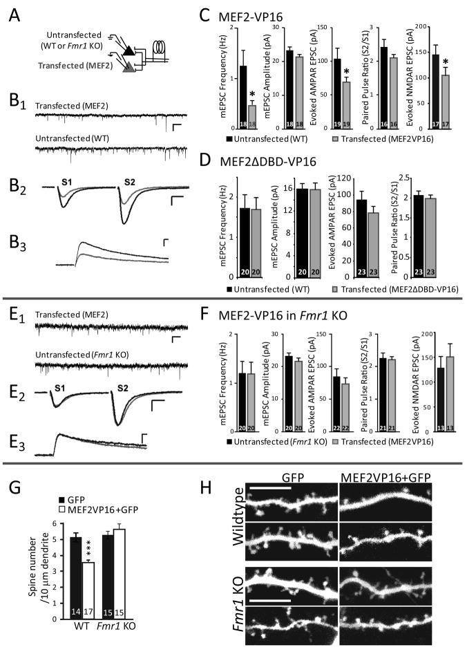
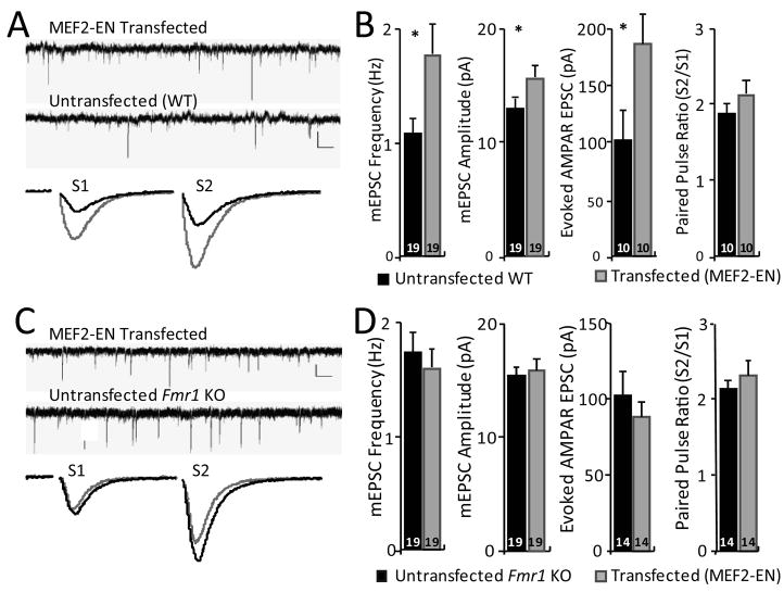
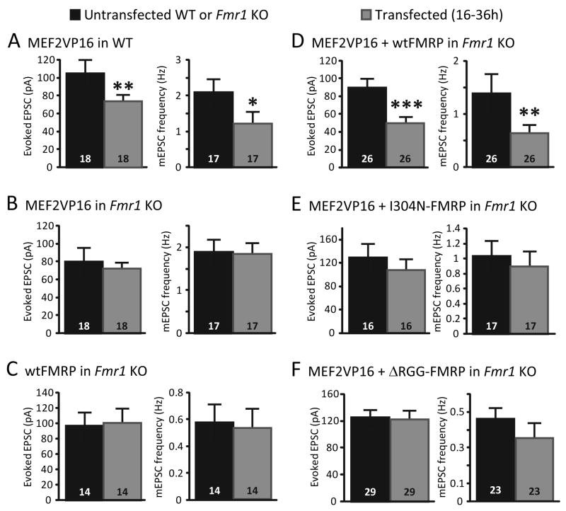
Fragile X syndrome (FXS), the most common genetic form of mental retardation and autism, is caused by loss-of-function mutations in an RNA-binding protein, Fragile X Mental Retardation Protein (FMRP). Neurons from patients and the mouse Fmr1 knockout (KO) model are characterized by an excess of dendritic spines, suggesting a deficit in excitatory synapse elimination. In response to neuronal activity, myocyte enhancer factor 2 (MEF2) transcription factors induce robust synapse elimination. Here, we demonstrate that MEF2 activation fails to eliminate functional or structural excitatory synapses in hippocampal neurons from Fmr1 KO mice. Similarly, inhibition of endogenous MEF2 increases synapse number in wild-type but not Fmr1 KO neurons. MEF2-dependent synapse elimination is rescued in Fmr1 KO neurons by acute postsynaptic expression of wild-type but not RNA-binding mutants of FMRP. Our results reveal that active MEF2 and FMRP function together in an acute, cell-autonomous mechanism to eliminate excitatory synapses.


Figures

Figure 1
Figure 1. MEF2 activation induces functional and structural synapse elimination in wildtype, but not Fmr1 KO neurons
A Experimental recording configuration. Organotypic hippocampal slice cultures from p6–7 wild-type (WT) or Fmr1 KO mice were biolistically transfected with MEF2-VP16-ER and treated with 4-hydroxytamoxifen (4-OHT) for 24–48 hours to induce MEF2-VP16-ER activation. Synaptic function was measured using dual simultaneous whole-cell patch clamp recordings of transfected and neighboring untransfected CA1 neurons. B1,B2, Representative traces of mEPSCs (B1; scale bar = 10 pA/500 ms.) and evoked AMPAR mediated EPSCs (B2; scale bar = 50 pA/10 ms) from a simultaneous recording from an untransfected and a neighboring MEF2-VP16-ER transfected WT neuron. B3 Pharmacologically isolated NMDAR mediated EPSCs from untransfected and neighboring MEF2-VP16 transfected WT neurons (scale bar = 50 pA/50 ms). C. Average mEPSC frequency, mEPSC amplitude, evoked AMPAR-mediated EPSC amplitude, and paired-pulse ratio (amplitude EPSC2/EPSC1) from untransfected WT and MEF2-VP16-ER transfected cells. Group data of pharmacologically isolated, evoked, NMDAR mediated EPSC amplitudes from untransfected and neighboring MEF2-VP16 transfected WT neurons. In this and all figures, averages are plotted + SEM and n (# of cell pairs) is indicated on each bar. * p< 0.05. D. Transfection of WT slice cultures with MEF2ΔDBD-VP16-ER, a DNA binding-deficient mutant of MEF2, and treatment with 4OHT does not alter excitatory synaptic transmission. Plotted are average mEPSC frequency, mEPSC amplitude, evoked EPSC amplitude, and paired-pulse ratio from untransfected and MEF2ΔDBD-VP16-ER transfected Fmr1 KO neurons. E, F. MEF2-VP16-ER transfection into Fmr1 KO slice cultures. E1, E2. Representative traces of mEPSCs (E1; scale bar = 10 pA/500 ms.) and evoked EPSCs (E2; scale bar = 50 pA/10 ms) from a simultaneous recording of an untransfected and neighboring MEF2-VP16-ER transfected Fmr1 KO neuron. E3 Pharmacologically isolated NMDAR mediated EPSCs from untransfected and neighboring MEF2-VP16 transfected Fmr1 KO neurons (scale bar = 50 pA/50 ms). F. Plotted are average mEPSC frequency, mEPSC amplitude, evoked AMPAR mediated EPSC amplitude, and paired-pulse ratio from untransfected and MEF2-VP16-ER transfected Fmr1 KO neurons. Group data of pharmacologically isolated, evoked, NMDAR mediated EPSC amplitudes from untransfected and neighboring MEF2-VP16 transfected Fmr1 KO neurons., G. MEF2-VP16 reduces average dendritic spine number of wildtype neurons, but not of Fmr1 KO neurons. *** p< 0.001. H. Representative images of apical dendrites from wildtype or Fmr1 KO CA1 pyramidal neurons in slice cultures transfected with GFP alone or MEF2-VP16 + GFP for 36 hours. Scale bar = 10 μm.
Figure 2
Figure 2. Inhibition of endogenous MEF2 function enhances synapse number in wildtype, but not in Fmr1 KO neurons
A, Representative traces of mEPSCs (upper; scale bar = 10 pA/500 ms.) and evoked EPSCs (lower; scale bar = 50 pA/10 ms) from a simultaneous recording from an untransfected and neighboring MEF2-Engrailed (MEF2-EN) transfected WT neuron. B. Average mEPSC frequency, mEPSC amplitude, evoked EPSC amplitude, and paired-pulse ratio from untransfected WT and MEF2-EN transfected cells. Averages are plotted + SEM and n (# of cell pairs) is indicated on each bar. * p< 0.05. C, D MEF2-EN transfection into Fmr1 KO slice cultures. C. Representative traces of mEPSCs (upper; scale bar = 10 pA/500 ms.) and evoked EPSCs (lower; scale bar = 50 pA/10 ms) from a simultaneous recording of an untransfected and neighboring MEF2-EN transfected Fmr1 KO neuron. D. Average mEPSC frequency, mEPSC amplitude, evoked EPSC amplitude, and paired-pulse ratio from untransfected and MEF2-EN transfected Fmr1 KO neurons.
Figure 3
Figure 3. MEF2 levels and MEF2-induced transcription are normal in Fmr1 KO hippocampal neurons
A Hippocampal lysates from WT and Fmr1 KO littermates (P12–14) were probed for the major MEF2 isoforms, MEF2A and MEF2D. B. Quantification of MEF2A and MEF2D levels (normalized to β-tubulin) WT and Fmr1 KO hippocampi (N = 6 mice from each genotype). C. Dissociated hippocampal neurons (7–8 days in vitro) from WT and Fmr1 KO mice were transfected with MEF2-VP16-ER, the MEF2 transcriptional reporter, MRE-GFP, and mCherry (transfection indicator), treated 16–24hr with vehicle or 4-OHT, then imaged live. Scale bar = 100 μm. D. Group data of tamoxifen induced MRE-GFP positive WT and Fmr1 KO neurons expressed as a ratio of total number of transfected (mCherry-positive) neurons (mean±SEM; *** p < 0.001). The number of cells per condition >200 for each condition, n = 3 cultures. E. Neuronal depolarization of hippocampal slice cultures from WT and Fmr1 KO was induced for the indicated times with high K+ (55 mM KCl), isotonic media which activates MEF2 mediated transcription (Flavell et al., 2006). Quantitative real time PCR reveals similar activity-dependent induction of the MEF2 transcript Nurr77 in WT and Fmr1 KO cultures (n = 3 cultures (mice) for each time point). F. PC12 cells were transfected with MEF2-VP16-ER and treated with tamoxifen for the indicated times. Endogenous mRNA levels of Nurr77 and Fmr1 were determined by quantitative RT_PCR. MEF2 activation of PC12 cells effectively induces Nurr77 transcription (F1), but not Fmr1 (F2), indicating that Fmr1 is not a target gene regulated by MEF2 activation over the 8 hour time period analyzed..
Figure 4
Figure 4. Acute postsynaptic coexpression of MEF2 and FMRP are required for functional synapse elimination
A. Acute transfection of constitutively-active MEF2 (MEF2-VP16) into wildtype (WT) hippocampal slice cultures results in a decrease in synaptic transmission 16- 30 hrs post-transfection as measured by evoked EPSCs and mEPSC frequency. B. In contrast, acute transfection of MEF2-VP16 into Fmr1 KO hippocampal slice cultures has no effect on any measure of synapse function. C. Transfection of Fmr1 KO slice cultures with wildtype (wt) FMRP-GFP alone for 16–48 hours had no effect on evoked EPSCs or mEPSC frequency. D. Co-transfection of wtFMRP-GFP together with MEF2-VP16 into Fmr1 KO neurons (16–30 hrs) reduced EPSC size and mEPSC frequency. E, F. Co-transfection of I304N FMRP (E) or ΔRGG-FMRP (F) together with MEF2-VP16 (16–30 hrs) had no effect on evoked EPSCs or mEPSCs. For all experiments (A-F) mEPSC amplitudes and paired-pulse facilitation of evoked EPSCs were unaffected by transfection. *p< 0.05; **p< 0.01; *** p < 0.001.

References

    1. Abrahams BS, Geschwind DH. Advances in autism genetics: on the threshold of a new neurobiology. Nat Rev Genet. 2008;9:341–355. - PMC - PubMed
    1. Arnold MA, Kim Y, Czubryt MP, Phan D, McAnally J, Qi X, Shelton JM, Richardson JA, Bassel-Duby R, Olson EN. MEF2C transcription factor controls chondrocyte hypertrophy and bone development. Dev Cell. 2007;12:377–389. - PubMed
    1. Barbosa AC, Kim MS, Ertunc M, Adachi M, Nelson ED, McAnally J, Richardson JA, Kavalali ET, Monteggia LM, Bassel-Duby R, Olson EN. MEF2C, a transcription factor that facilitates learning and memory by negative regulation of synapse numbers and function. Proc Natl Acad Sci U S A. 2008;105:9391–9396. - PMC - PubMed
    1. Bassell GJ, Warren ST. Fragile X syndrome: loss of local mRNA regulation alters synaptic development and function. Neuron. 2008;60:201–214. - PMC - PubMed
    1. Busetto G, Higley MJ, Sabatini BL. Developmental presence and disappearance of postsynaptically silent synapses on dendritic spines of rat layer 2/3 pyramidal neurons. J Physiol. 2008;586:1519–1527. - PMC - PubMed

Publication types

MeSH terms

Substances