

Use of specific glycosidases to probe cellular interactions in the sea urchin embryo
- PMID: 20435035
- PMCID: PMC2921930
- DOI: 10.1016/j.yexcr.2010.04.026
Use of specific glycosidases to probe cellular interactions in the sea urchin embryo
Abstract
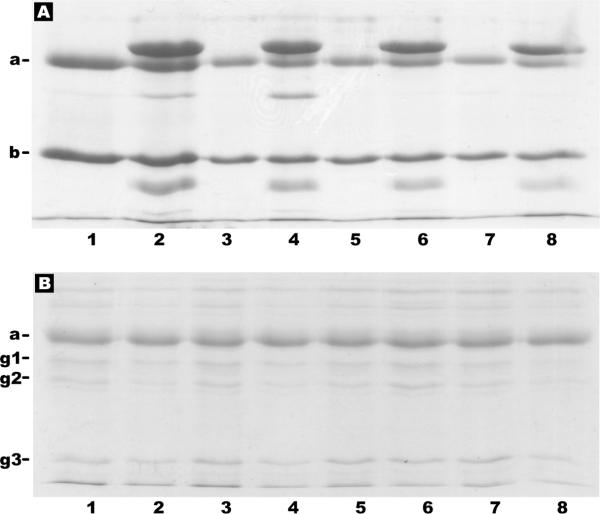
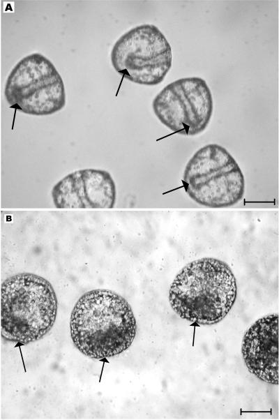
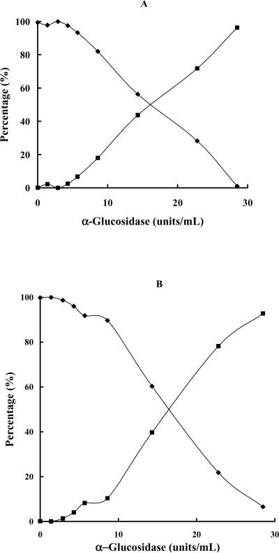
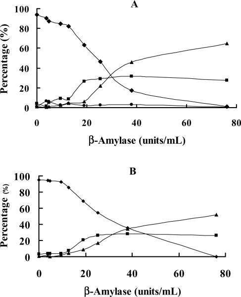
We present an unusual and novel model for initial investigations of a putative role for specifically conformed glycans in cellular interactions. We have used alpha- and ss-amylase and alpha- and ss-glucosidase in dose-response experiments evaluating their effects on archenteron organization using the NIH designated sea urchin embryo model. In quantitative dose-response experiments, we show that defined activity levels of alpha-glucosidase and ss-amylase inhibited archenteron organization in living Lytechinus pictus gastrula embryos, whereas all concentrations of ss-glucosidase and alpha-amylase were without substantial effects on development. Product inhibition studies suggested that the enzymes were acting by their specific glycosidase activities and polyacrylamide gel electrophoresis suggested that there was no detectable protease contamination in the active enzyme samples. The results provide evidence for a role of glycans in sea urchin embryo cellular interactions with special reference to the possible structural conformation of these glycans based on the differential activities of the alpha- and ss-glycosidases.
Copyright 2010 Elsevier Inc. All rights reserved.
Figures
Similar articles
-
Carbohydrate involvement in cellular interactions in sea urchin gastrulation.Acta Histochem. 2004;106(2):97-106. doi: 10.1016/j.acthis.2004.01.001. Acta Histochem. 2004. PMID: 15147630
-
A role for polyglucans in a model sea urchin embryo cellular interaction.Zygote. 2014 Aug;22(3):419-29. doi: 10.1017/S0967199413000038. Epub 2013 Mar 27. Zygote. 2014. PMID: 23534855
-
Terminal alpha-d-mannosides are critical during sea urchin gastrulation.Zygote. 2016 Oct;24(5):775-82. doi: 10.1017/S0967199416000113. Epub 2016 May 18. Zygote. 2016. PMID: 27189235
-
The painted sea urchin, Lytechinus pictus, as a genetically-enabled developmental model.Methods Cell Biol. 2019;150:105-123. doi: 10.1016/bs.mcb.2018.11.010. Epub 2019 Feb 10. Methods Cell Biol. 2019. PMID: 30777173 Free PMC article. Review.
-
Gene regulatory networks for ectoderm specification in sea urchin embryos.Biochim Biophys Acta. 2009 Apr;1789(4):261-7. doi: 10.1016/j.bbagrm.2009.02.002. Epub 2009 Mar 21. Biochim Biophys Acta. 2009. PMID: 19429544 Review.
Cited by
-
Glyconectin Cell Adhesion Epitope, β-d-GlcpNAc3S-(1→3)-α-l-Fucp, Is Involved in Blastulation of Lytechinus pictus Sea Urchin Embryos.Molecules. 2021 Jun 30;26(13):4012. doi: 10.3390/molecules26134012. Molecules. 2021. PMID: 34209220 Free PMC article.
References
-
- Ernst SG. A century of sea urchin development. American Zoologist. 1997;37:250–250.
-
- Davidson EH. The sea urchin genome: where will it lead us? Science. 2006;314:939–940. - PubMed
-
- E.H., C. Davidson RA. Arguments for sequencing the genome of the sea urchin Strongylocentrotus purpuratus. 2002. - PubMed
-
- Oppenheimer SB, Carroll EJ., Jr. Introduction to Embryonic Development, Pearson Education, Upper Saddle River. 2004.
Publication types
MeSH terms
Substances
Grants and funding
LinkOut - more resources
Full Text Sources
Miscellaneous
