. 2010 Oct;126(2):198-202.
doi: 10.1016/j.exppara.2010.04.022. Epub 2010 May 18.

Cloning of Plasmodium falciparum by single-cell sorting

Affiliations

Cloning of Plasmodium falciparum by single-cell sorting

Jun Miao et al. Exp Parasitol. 2010 Oct.

Abstract

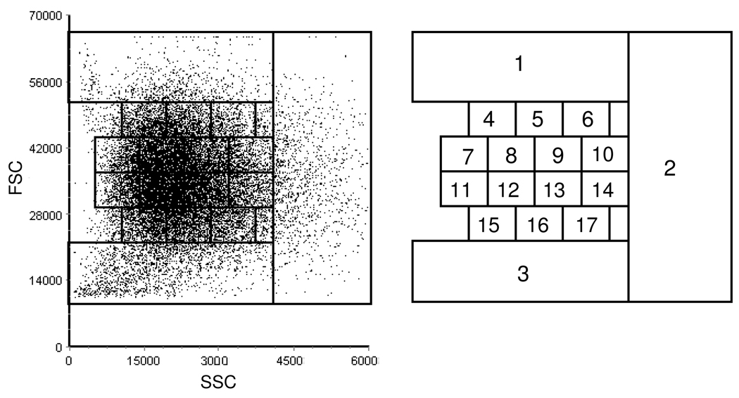
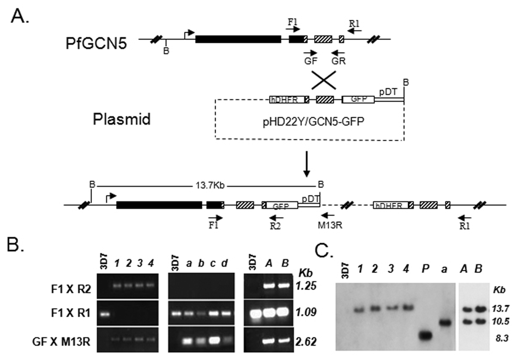
Malaria parasite cloning is traditionally carried out mainly by using the limiting dilution method, which is laborious, imprecise, and unable to distinguish multiply-infected RBCs. In this study, we used a parasite engineered to express green fluorescent protein (GFP) to evaluate a single-cell sorting method for rapidly cloning Plasmodium falciparum. By dividing a two-dimensional scattergram from a cell sorter into 17 gates, we determined the parameters for isolating singly-infected erythrocytes and sorted them into individual cultures. Pre-gating of the engineered parasites for GFP allowed the isolation of almost 100% GFP-positive clones. Compared with the limiting dilution method, the number of parasite clones obtained by single-cell sorting was much higher. Molecular analyses showed that parasite isolates obtained by single-cell sorting were highly homogenous. This highly efficient single-cell sorting method should prove very useful for cloning both P. falciparum laboratory populations from genetic manipulation experiments and clinical samples.


Figures

Fig. 1
Fig. 1. Scattergram of infected RBCs and determination of FSC and SSC parameters for isolating singly-infected RBCs
Purified trophozoites were subjected to cell sorting, and the FSC and SSC scattergram was shown in the left panel. From this scatter pattern, 17 gates were set to cover the plotted area (right panel). 106 particles from each gate were sorted into individual tubes, made into thin smears, and analyzed by Giemsa staining. Gates 12 and 13 contained mostly singly-infected RBCs.
Fig. 2
Fig. 2. Tagging of the C-terminus of PfGCN5 with GFP and characterization of clones
(A) Predicted GFP integration from a single-crossover event. Restriction enzyme BamHI sites (B) and the expected sizes of DNA fragments after BamHI digestion are shown. Top: the PfGCN5 locus on chromosome 11. Solid lines indicate introns or intergenic regions, filled boxes are exons, and hatched boxes are the C-terminal end for homologous recombination. Middle: The transfection plasmid pHD22Y/GCN5-GFP showing the PfGCN5 genomic fragment fusion with GFP, pDT 3’, and the drug selection cassette hDHFR. Bottom: The predicted single-crossover event at the PfGCN5 locus showing the integration of one copy of the plasmid. The primers F1 × R2, GF × M13R and F1 × R1 were designed for identifying this integration event, confirmation of the presence of the transfected plasmid, and detecting the intact PfGCN5 locus, respectively. (B) Confirmation of chromosomal integration and clones by PCR. Genomic DNA of wild-type 3D7 and clones were amplified with primers F1 × R2, F1 × R1 and GF × M13R, and the sizes of the PCR products are indicated. All clones (except the wild type 3D7) were positive for GF × M13R, indicating the presence of the transfection plasmid in the parasites. Four clones (1, 2, 3 and 4) showed PCR amplification with F1 × R2, but not F1 × R1, suggesting of GFP integration at the PfGCN5 locus and no contamination with parasites with other integration events. Four other clones (a, b, c and d) showed the opposite - negative with F1 × R2, but positive with F1 × R1, suggesting that the PfGCN5 was intact and plasmid was integrated elsewhere. Two wells (A and B) derived from limited dilution method showed PCR products for all three pairs of primers, suggesting that these parasites were not clonal and contained a mixture of parasites with GFP integrated at the PfGCN5 locus and elsewhere in the genome (C) Verification of the predicted integration events and resulting clones by Southern blot. Genomic DNA from wild type 3D7, four clones with integration at the PfGCN5 locus (1, 2, 3 and 4), one clone with plasmid integrated at another locus of the genome (a), 100 ng of the plasmid (P), and two isolates containing mixed clones (A and B) was digested with BamH1, separated in a 0.6% agarose gel, and hybridized to 32P-labeled GFP PCR product. Integration at the PfGCN5 locus only showed a 13.7 Kb band, whereas integration of the plasmid at another locus had a 10.5 kb band. Two bands were observed in isolates A and B, suggesting of mixed clones.
Fig. 3
Fig. 3. Pre-gating of GFP-expressing parasites before single-cell sorting
(A) GFP expressing trophozoites were selected by setting a gate (M1). Parasites were scanned with the GFP filter to measure the GFP fluorescence intensity of each parasite. Parasites with GFP signal intensity falling within the M1 gate were selected for single-cell sorting. (B) GFP images of single clones from single-cell cloning were captured under a fluorescence microscope.

References

    1. Allen RJ, Kirk K. Plasmodium falciparum culture: The benefits of shaking. Molecular and Biochemical Parasitology. 2009;169:63–65. - PubMed
    1. Barkan D, Ginsburg H, Golenser J. Optimisation of flow cytometric measurement of parasitaemia in plasmodium-infected mice. International Journal for Parasitology. 2000;30:649–653. - PubMed
    1. Bhakdi SC, Sratongno P, Chimma P, Rungruang T, Chuncharunee A, Neumann HP, Malasit P, Pattanapanyasat K. Re-evaluating acridine orange for rapid flow cytometric enumeration of parasitemia in malaria-infected rodents. Cytometry A. 2007;71:662–667. - PubMed
    1. Chen Q, Fernandez V, Sundstrom A, Schlichtherle M, Datta S, Hagblom P, Wahlgren M. Developmental selection of var gene expression in Plasmodium falciparum. Nature. 1998;394:392–395. - PubMed
    1. Cui L, Fan Q, Li J. The malaria parasite Plasmodium falciparum encodes members of the Puf RNA-binding protein family with conserved RNA binding activity. Nucleic Acids Research. 2002;30:4607–4617. - PMC - PubMed

Publication types

MeSH terms

Substances