. 2010 Aug 11;169(1):182-94.
doi: 10.1016/j.neuroscience.2010.04.056. Epub 2010 May 7.

Dopamine receptor expression and distribution dynamically change in the rat nucleus accumbens after withdrawal from cocaine self-administration

Affiliations

Dopamine receptor expression and distribution dynamically change in the rat nucleus accumbens after withdrawal from cocaine self-administration

K L Conrad et al. Neuroscience. .

Abstract

Dopamine receptors (DARs) in the nucleus accumbens (NAc) are critical for cocaine's actions but the nature of adaptations in DAR function after repeated cocaine exposure remains controversial. This may be due in part to the fact that different methods used in previous studies measured different DAR pools. In the present study, we used a protein crosslinking assay to make the first measurements of DAR surface expression in the NAc of cocaine-experienced rats. Intracellular and total receptor levels were also quantified. Rats self-administered saline or cocaine for 10 days. The entire NAc, or core and shell subregions, were collected one or 45 days later, when rats are known to exhibit low and high levels of cue-induced drug seeking, respectively. We found increased cell surface D1 DARs in the NAc shell on the first day after discontinuing cocaine self-administration (designated withdrawal day 1, or WD1) but this normalized by WD45. Decreased intracellular and surface D2 DAR levels were observed in the cocaine group. In shell, both measures decreased on WD1 and WD45. In core, decreased D2 DAR surface expression was only observed on WD45. Similarly, WD45 but not WD1 was associated with increased D3 DAR surface expression in the core. Taking into account many other studies, we suggest that decreased D2 DAR and increased D3 DAR surface expression on WD45 may contribute to enhanced cocaine-seeking after prolonged withdrawal, although this is likely to be a modulatory effect, in light of the mediating effect previously demonstrated for AMPA-type glutamate receptors.


Figures

Fig. 1
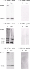
Fig. 1. Measurement of DAR surface expression using a protein crosslinking assay and demonstration of immunospecificity by preabsorbing DAR antibodies with peptides used to raise each antibody
The membrane impermeant protein crosslinking reagent BS3 crosslinks surface-expressed proteins, forming high molecular weight aggregates, but does not modify intracellular proteins. Surface and intracellular DAR can therefore be distinguished by molecular weight. (A-C) Crosslinked (X) and non-crosslinked (Non) NAc tissue (20-30μg protein/lane) was probed with antibodies to the D1, D2, and D3 DARs, respectively, in the presence of the vehicle (TBS) used to dissolve the peptides (DAR ab + Veh groups). A surface band (>250 kDa) was evident only in the crosslinked tissue. A single intracellular band was observed for the D1 DAR (A) and D3 DAR (C). For the D2 DAR (B), three intracellular bands were observed, presumably corresponding to the D2 DAR monomer (~55kDa), glycosylated monomer (~75kDa), and dimer (~100kDa). (D-F) Preabsorption of the D1, D2, or D3 DAR antibodies, respectively, with the peptide used to raise each antibody nearly eliminated all surface and intracellular bands (DAR ab + peptide groups). For each of the DAR, we quantified diffuse density of bands in arbitrary units. As expected from prior studies on AMPAR (Boudreau and Wolf, 2005), we found that the sum of surface (S) and intracellular (I) bands in the crosslinked lanes approximately equaled the total (T) DAR protein in the non-crosslinked lanes (i.e., S + I summed to 92.47% of T for the D1 DAR, 109.15% for the D2 DAR, and 98.9% for the D3 DAR).
Fig. 2
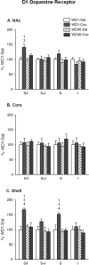
Fig. 2. D1 DAR surface expression was increased in the NAc shell after 1 day of withdrawal from cocaine self-administration
Surface (I), intracellular (I), and total (S+I) D1 DAR levels, and the surface/intracellular ratio (S/I), were determined in the entire NAc (A), the core subregion (B), and the shell subregion (C). Data (mean ± S.E.M, NAc: n = 7-12 per group; NAc core and shell: n = 5-7 per group) are expressed as a percentage of the WD1-Sal group. S, I and S+I values were normalized to total protein in the lane; the S/I ratio is independent of total protein loaded onto the gel. 1p < 0.05 compared with WD1-Sal; 2p < 0.05 compared with WD1-Coc; 3p < 0.05 compared with WD45-Sal; 4p < 0.05 compared with WD45-Coc.
Fig. 3
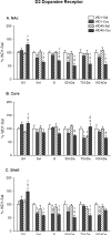
Fig. 3. Intracellular and surface D2 DAR levels in the NAc were decreased after 45 days of withdrawal from cocaine self-administration
Surface (I), intracellular (I), and total (S+I) D2 DAR levels, and the surface/intracellular ratio (S/I), were determined in the entire NAc (A), the core subregion (B), and the shell subregion (C). For calculations of S+I and S/I, all three intracellular bands (~55, 75 and 100 kDa; see Figure 1) were summed to generate I. Data (mean ± S.E.M, NAc: n = 7-12 per group; NAc core and shell: n = 5-7 per group) are expressed as a percentage of the WD1-Sal group. S, I and S+I values were normalized to total protein in the lane; S/I is independent of total protein loaded onto the gel. 1p < 0.05 compared with WD1-Sal; 2p < 0.05 compared with WD1-Coc; 3p < 0.05 compared with WD45-Sal; 4p < 0.05 compared with WD45-Coc.
Fig. 4
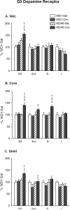
Fig. 4. D3 DAR surface expression was increased in the NAc after 45 days of withdrawal from cocaine self-administration
Surface (I), intracellular (I), and total (S+I) D3 DAR levels, and the surface/intracellular ratio (S/I), were determined in the entire NAc (A), the core subregion (B), and the shell subregion (C). Data (mean ± S.E.M, NAc: n = 7-12 per group; NAc core and shell: n = 5-7 per group) are expressed as a percentage of the WD1-Sal group. S, I and S+I values were normalized to total protein in the lane; S/I is independent of total protein loaded onto the gel. 1p < 0.05 compared with WD1-Sal; 2p < 0.05 compared with WD1-Coc; 3p < 0.05 compared with WD45-Sal; 4p < 0.05 compared with WD45-Coc.

References

    1. Alleweireldt AT, Hobbs RJ, Taylor AR, Neisewander JL. Effects of SCH-23390 infused into the amygdala or adjacent cortex and basal ganglia on cocaine seeking and self-administration in rats. Neuropsychopharmacology. 2006;31:363–374. - PubMed
    1. Anderson SM, Bari AA, Pierce RC. Administration of the D1-like dopamine receptor antagonist SCH-23390 into the medial nucleus accumbens shell attenuates cocaine priming-induced reinstatement of drug-seeking behavior in rats. Psychopharmacology (Berl) 2003;168:132–138. - PubMed
    1. Anderson SM, Famous KR, Sadri-Vakili G, Kumaresan V, Schmidt HD, Bass CE, Terwilliger EF, Cha JH, Pierce RC. CaMKII: a biochemical bridge linking accumbens dopamine and glutamate systems in cocaine seeking. Nat Neurosci. 2008;11:344–353. - PubMed
    1. Anderson SM, Pierce RC. Cocaine-induced alterations in dopamine receptor signaling: implications for reinforcement and reinstatement. Pharmacol and Ther. 2005;106:389–403. - PubMed
    1. Bachtell RK, Whisler K, Karanian D, Self DW. Effects of intra-nucleus accumbens shell administration of dopamine agonists and antagonists on cocaine-taking and cocaine-seeking behaviors in the rat. Psychopharmacology (Berl) 2005;183:41–53. - PubMed

Publication types

MeSH terms