. 2010 Jul;299(1):H193-201.
doi: 10.1152/ajpheart.00431.2009. Epub 2010 Apr 30.

Resistin decreases expression of endothelial nitric oxide synthase through oxidative stress in human coronary artery endothelial cells

Affiliations

Resistin decreases expression of endothelial nitric oxide synthase through oxidative stress in human coronary artery endothelial cells

Changyi Chen et al. Am J Physiol Heart Circ Physiol. 2010 Jul.

Abstract

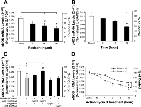
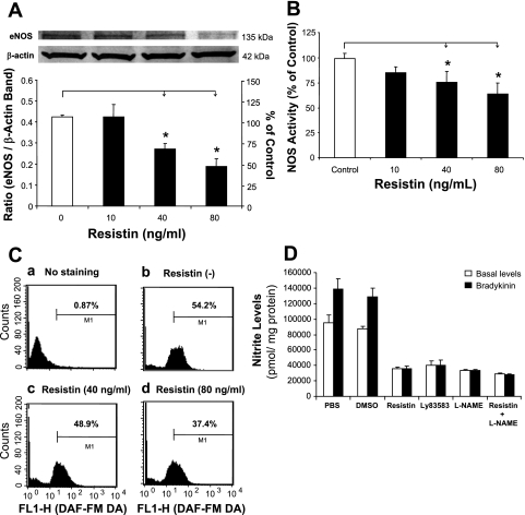
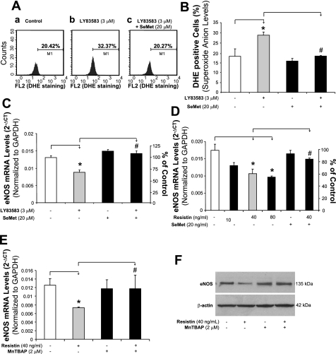
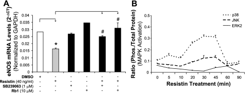
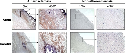
Resistin is a newly discovered adipocyte-derived cytokine that may play an important role in insulin resistance, diabetes, adipogenesis, inflammation, and cardiovascular disease. However, it is largely unknown whether resistin impairs endothelial functions by affecting the endothelial nitric oxide synthase (eNOS) system. In this study, we determined the effect of human recombinant resistin protein on eNOS expression and regulation in human coronary artery endothelial cells (HCAECs). When cells were treated with clinically relevant concentrations of resistin (40 or 80 ng/ml) for 24 h, the levels of eNOS mRNA, protein, and activity and eNOS mRNA stability were significantly reduced. Cellular nitric oxide levels were also decreased. In addition, the cellular levels of reactive oxygen species (ROS), including superoxide anion, were significantly increased in resistin-treated HCAECs. Mitochondrial membrane potential and the activities of catalase and superoxide dismutase were reduced. Three antioxidants, seleno-L-methionine, ginsenoside Rb1, and MnTBAP (superoxide dismutase mimetic), effectively blocked resistin-induced eNOS downregulation. Meanwhile, resistin activated the mitogen-activated protein kinases p38 and c-Jun NH(2)-terminal kinase (JNK), and the specific p38 inhibitor SB-239063 effectively blocked resistin-induced ROS production and eNOS downregulation. Furthermore, immunoreactivity of resistin was increased in atherosclerotic regions of human aorta and carotid arteries. Thus resistin directly induces eNOS downregulation through overproduction of ROS and activation of p38 and JNK in HCAECs. Resistin-induced mitochondrial dysfunction and imbalance in cellular redox enzymes may be the underlying mechanisms of oxidative stress.


Figures

Fig. 1.
Fig. 1.
Effect of resistin on endothelial nitric oxide synthase (eNOS) mRNA levels in human coronary artery endothelial cells (HCAECs). HCAECs were cultured with different concentrations of resistin for different periods of time. The mRNA levels of eNOS and glyceraldehyde-3-phosphate dehydrogenase (GAPDH) were determined by real-time PCR analysis. A: cells were treated with different concentrations of resistin (10, 40, or 80 ng/ml) for 24 h. *P < 0.05, n = 3 experiments. B: cells were treated with resistin (40 ng/ml) for different times (6, 12, and 24 h). *P < 0.05, n = 3. C: cells were treated with 40 ng/ml resistin and different antibodies (Ab) for 24 h. *P < 0.05, n = 3 (compared with untreated controls). #P < 0.05, n = 3 (compared with resistin treatment alone). D: HCAEC eNOS mRNA stability was determined by real-time PCR after cells were treated with 5 μg/ml actinomycin D in the presence or absence of resistin (40 ng/ml) for indicated time points (0, 0.5, 1, 3, 6, or 12 h). *P < 0.05, n = 3 experiments.
Fig. 2.
Fig. 2.
Effects of resistin on eNOS protein levels and NOS activity in HCAECs. A: Western blot analysis. HCAECs were treated with 40 or 80 ng/ml resistin for 24 h. Representative bands of eNOS and β-actin staining and quantitation of band density ratios (eNOS and β-actin). *P < 0.05, n = 3. B: NOS activity assay by fluorescence microplate reader. *P < 0.05, n = 3. C: flow cytometry (cellular NO levels). Treated HCAECs were stained with 10 μM of 4-amino-5-methylamino-2′,7′-difluorofluorescein diacetate (DAF-FM DA) for 30 min. D: nitric oxide (NO) levels in the supernatant of cell culture (Griess assay). HCAECs were treated with resistin (40 ng/ml) and/or other molecules [3 μM LY-83583 and 100 μM NG-nitro-l-arginine methyl ester (l-NAME)] for 24 h. Basal and bradykinin-stimulated levels of NO-derived nitrite in the culture supernatant were detected. *P < 0.05, n = 3.
Fig. 3.
Fig. 3.
Effect of resistin on oxidative stress in HCAECs. A: reactive oxygen species (ROS) production [dihydroethidium (DHE) staining and flow cytometry analysis]. HCAECs were treated with 40 or 80 ng/ml of resistin for 24 h. *P < 0.05, n = 3. B: superoxide anion (O2) levels (DHE staining and HPLC analysis). HCAECs were treated with resistin (40 ng/ml) for 3 h, and then DHE (25 μM) was added to the cells for 30 min. Cellular levels of 2-hydroxyethidium (2-EOH) and ethidium were determined by HPLC analysis. Positive control of 2-EOH was prepared by using nitrosodisulfonate radical dianion (NDS) with DHE in the aqueous phosphate buffer, pH 7.4, containing diethylene triamine pentaacetic acid (DTPA). Specific O2 scavenger manganese [III] tetrakis(4-benzoic acid)porphyrin [MnTBAP, superoxide dismutase (SOD) mimetic] was used. C: ROS production (glutathione assay). HCAECs were treated with resistin and/or other molecules for 24 h. RLU, relative light units; TTFA, thenoyltrifluoroacetone. *P < 0.05, n = 3 [compared with dimethyl sulfoxide (DMSO) controls]. #P < 0.05, n = 3 (compared with resistin treatment alone). D: time course of ROS production (glutathione assay). HCAECs were treated with resistin (40 μg/ml) for different time points. *P < 0.05, n = 3 [compared with dimethyl sulfoxide (DMSO) controls]. #P < 0.05, n = 3 (compared with resistin treatment alone). E: mitochondrial membrane potential (JC-1 staining and flow cytometry). F: catalase activity assay. HCAECs were treated with resistin (40 or 80 ng/ml) and/or selenomethionine (SeMet, 20 μM) for 24 h. *P < 0.05, n = 3 (compared with DMSO controls). #P < 0.05, n = 3 (compared with resistin treatment alone). G: SOD activity assay. HCAECs were treated with resistin (40 or 80 ng/ml) and/or SeMet (20 μM) for 24 h. *P < 0.05, n = 3 (compared with DMSO controls). #P < 0.05, n = 3 (compared with resistin treatment alone).
Fig. 4.
Fig. 4.
Effects of superoxide donor LY-83583, resistin, SeMet, and MnTBAP on eNOS mRNA expression in HCAECs. A: ROS production (DHE staining and flow cytometry analysis). HCAECs were treated with LY-83583 (3 μM) and/or SeMet (20 μM) for 24 h. B: quantitative data of LY-83583-induced ROS overproduction. *P < 0.05, n = 3 (compared with DMSO controls). #P < 0.05, n = 3 (compared with resistin treatment alone). C: eNOS mRNA (real-time PCR assay) in LY-83583-treated HCAECs. *P < 0.05, n = 3 (compared with DMSO controls). #P < 0.05, n = 3 (compared with resistin treatment alone). D: effect of SeMet on eNOS mRNA levels (real-time PCR assay) in HCAECs. *P < 0.05, n = 3 (compared with DMSO controls). #P < 0.05, n = 3 (compared with resistin treatment alone). E: effect of MnTBAP on eNOS mRNA levels (real-time PCR assay) in HCAECs. *P < 0.05, n = 3 (compared with DMSO controls). #P < 0.05, n = 3 (compared with resistin treatment alone). F: effect of MnTBAP on eNOS protein levels (Western blot analysis) in HCAECs.
Fig. 5.
Fig. 5.
Role of mitogen-activated protein kinases (MAPKs) and ginsenoside Rb1 on resistin-induced eNOS downregulation. A: eNOS mRNA levels (real-time PCR assay). HCAECs were treated with resistin and/or other molecules (p38 inhibitor SB-239063 and ginsenoside Rb1) for 24 h. *P < 0.05, n = 3 (compared with DMSO controls). #P < 0.05, n = 3 (compared with resistin treatment alone). B: phosphorylation of MAPKs [p38, c-Jun NH2-terminal kinase (JNK), and extracellular signal-regulated kinase (ERK) 2] by using Bio-Rad Bioplex luminex immunoassay. HCAECs were treated with resistin (40 ng/ml) for different time points.
Fig. 6.
Fig. 6.
Resistin immunoreactivity of human atherosclerotic and nonatherosclerotic arteries. Human aorta and carotid arteries were fixed in formalin and embedded in paraffin. Immunostaining was performed by using the anti-human resistin antibody (1:200), biotinylated secondary antibody, and avidin-biotin reaction using peroxidase enzyme. Brown color represents positive staining of resistin.

References

    1. Allan CB, Lacourciere GM, Stadtman TC. Responsiveness of selenoproteins to dietary selenium. Annu Rev Nutr 19: 1–16, 1999 - PubMed
    1. Azumi H, Inoue N, Ohashi Y, Terashima M, Mori T, Fujita H, Awano K, Kobayashi K, Maeda K, Hata K, Shinke T, Kobayashi S, Hirata K, Kawashima S, Itabe H, Hayashi Y, Imajoh-Ohmi S, Itoh H, Yokoyama M. Superoxide generation in directional coronary atherectomy specimens of patients with angina pectoris: important role of NAD(P)H oxidase. Arterioscler Thromb Vasc Biol 22: 1838–1844, 2002 - PubMed
    1. Battin EE, Perron NR, Brumaghim JL. The central role of metal coordination in selenium antioxidant activity. Inorg Chem 45: 499–501, 2006 - PubMed
    1. Burnett MS, Lee CW, Kinnaird TD, Stabile E, Durrani S, Dullum MK, Devaney JM, Fishman C, Stamou S, Canos D, Zbinden S, Clavijo LC, Jang GJ, Andrews JA, Zhu J, Epstein SE. The potential role of resistin in atherogenesis. Atherosclerosis 182: 241–248, 2005 - PubMed
    1. Calabro P, Yeh ET. Intra-abdominal adiposity, inflammation, and cardiovascular risk: new insight into global cardiometabolic risk. Curr Hypertens Rep 10: 32–38, 2008 - PubMed

Publication types

MeSH terms