. 2010 Jul 15;116(2):287-96.
doi: 10.1182/blood-2009-12-259598. Epub 2010 Apr 30.

SOCS3 regulates graft-versus-host disease

Affiliations

SOCS3 regulates graft-versus-host disease

Geoffrey R Hill et al. Blood. .

Abstract

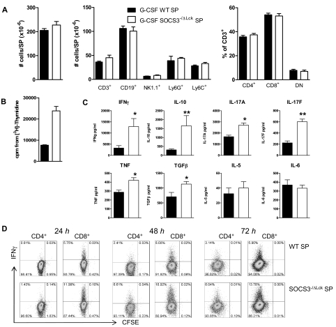
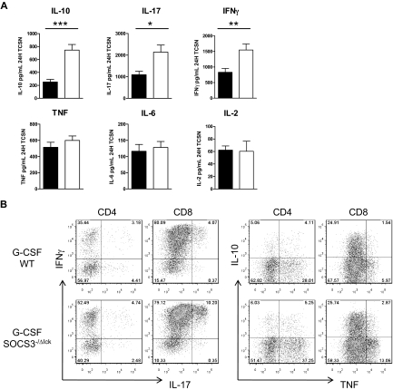
Suppressor of cytokine signaling-3 (SOCS3) is the main intracellular regulator of signaling by granulocyte colony-stimulating factor, an immune-modulatory cytokine used to mobilize stem cells for transplantation. We have therefore studied the contribution of SOCS3 to the spectrum of graft-versus-host disease (GVHD) after allogeneic stem cell transplantation (SCT). Grafts from SOCS3(-/Deltavav) donor mice in which SOCS3 deficiency is restricted to the hematopoietic compartment had an augmented capacity to induce acute GVHD. With the use of SOCS3(-/DeltaLysM) and SOCS3(-/Deltalck) donors in which SOCS3 deficiency was restricted to the myeloid or T-cell lineage, respectively, we confirmed SOCS3 deficiency promoted acute GVHD mortality and histopathology within the gastrointestinal tract by effects solely within the donor T cell. SOCS3(-/Deltalck) donor T cells underwent enhanced alloantigen-dependent proliferation and generation of interleukin-10 (IL-10), IL-17, and interferon-gamma (IFNgamma) after SCT. The enhanced capacity of the SOCS3(-/Deltalck) donor T cell to induce acute GVHD was dependent on IFNgamma but independent of IL-10 or IL-17. Surprisingly, SOCS3(-/Deltalck) donor T cells also induced severe, transforming growth factor beta- and IFNgamma-dependent, sclerodermatous GVHD. Thus, the delivery of small molecule SOCS3 mimetics may prove to be useful for the inhibition of both acute and chronic GVHD.


Figures

Figure 1
Figure 1
SOCS3 within donor T cells attenuates GVHD after G-CSF–mobilized allogeneic SCT. Donor B6 mice were treated with G-CSF (10 μg/animal per day for 4 days) or were untreated. Unfractionated splenic grafts containing 2 million T cells were transplanted into lethally irradiated (1100 cGy) B6D2F1 recipient mice. Survival curves by Kaplan-Meier analysis, pooled from 2 experiments. ***P < .001 for recipients of G-CSF Socs3−/Δlck versus G-CSF WT spleen; *P < .05 for recipients of G-CSF SOCS3−/Δvav versus G-CSF WT spleen.
Figure 2
Figure 2
SOCS3−/ΔLck T cells are hyperresponsive to T-cell receptor ligation after G-CSF mobilization. (A) Size and cell subset composition of spleens from G-CSF–treated WT or SOCS3−/ΔLck mice. Data are representative of 3 similar experiments (n = 3 animals/group). DN indicates double negative. (B) Splenocytes from G-CSF–treated WT or SOCS3−/ΔLck mice were cultured in the presence of soluble CD3, and proliferative responses were assessed by [3H] thymidine incorporation. (C) Supernatants from CD3 cultures were harvested at 72 hours and assayed for cytokines by cytometric bead array or enzyme-linked immunoabsorbent assay (IL-17A, IL-17F, and TGFβ). Data are pooled from 2 independent experiments, n = 4-6 animals/group. *P < .05; ** P < .01. (D) IFNγ secretion by purified CFSE-labeled T cells at the times indicated after stimulation with plate-bound CD3 and CD28. Data are representative of 2 experiments.
Figure 3
Figure 3
SOCS3 within T cells limits T-cell proliferation and apoptosis after allogeneic SCT. Splenocytes from G-CSF–treated WT or SOCS3−/ΔLck mice were CFSE labeled, and grafts containing 2 million T cells were transplanted into lethally irradiated (1100 cGy) B6D2F1 recipient mice. (A) Spleens were harvested from recipients 3 days later, and CFSE dilution in the CD4+ T-cell compartment was examined by flow cytometry. (B) Modfit CFSE dilution analysis of splenic and lymph node T-cell proliferation. Data are representative of 2 similar experiments with 4 to 5 animals/group. (C) Splenocytes from G-CSF–treated WT or SOCS3−/ΔLck mice were transplanted into lethally irradiated (1100 cGy) B6D2F1 recipient mice. On days 7 and 14 after transplantation, spleens were harvested, total cellularity was determined with an automated cell counter, and CD4+ and CD8+ T cells were quantified by flow cytometry. (D) On day 7 after transplantation, spleens were stained with annexin V and 7-amino-actinomycin (7AAD), and the frequency of apoptotic splenocytes (annexin V+7AAD) was determined by flow cytometric analysis. Black bars and white bars represent G-CS–treated WT or SOCS3−/ΔLck mice, respectively.
Figure 4
Figure 4
SOCS3−/ΔLck T cells exhibit dysregulated cytokine production after allogeneic SCT. Splenocytes from G-CSF–treated WT or SOCS3−/ΔLck mice were transplanted into lethally irradiated (1100 cGy) B6D2F1 recipient mice, and at day 7 after transplantation splenocytes were examined for cytokine production. (A) Splenocytes were cultured for 24 hours with soluble CD3, and supernatants were collected and assayed for cytokines by cytometric bead array. Data represent mean ± SEM of pooled results from 2 similar experiments (n = 10 animals/group). Black bars and white bars represent G-CSF–treated WT or SOCS3−/ΔLck mice, respectively. (B) After 4-hour culture with phorbol myristate acetate and ionomycin, cell cytokine production was analyzed by intracellular cytokine staining with 4-color flow cytometry. Numbers in quadrants represent the percentage of gated CD4 or CD8 T cells as indicated.
Figure 5
Figure 5
Enhanced IFNγ generation by SOCS3−/ΔLck T cells drives gastrointestinal tissue destruction after allogeneic SCT. SOCS3−/ΔLck donors were treated with G-CSF, and unfractionated splenocytes containing 2 million T cells or T cell–depleted grafts were transplanted into lethally irradiated B6D2F1 recipient mice. Survival curves by Kaplan-Meier analysis. (A) Transplant recipients received isotype control mAb or anti–IL-10R thrice/week from day 7 onward. Data represent a single experiment with 10 animals/GVHD group, and 4 animals/TCD group. (B) Transplant recipients received isotype control mAb or anti–IL-17 mAb thrice/week from day 0 onward. Data are pooled from 3 similar experiments (n = 24 animals/GVHD group, n = 12 animals/TCD group). (C) Transplant recipients received T cell–replete or T cell–depleted grafts from G-CSF–treated WT or SOCS3−/ΔLck donors, and small bowel, liver, and skin histopathology were assessed at day 7 after transplantation as described in “Histology.” (D) Images of hematoxylin and eosin–stained sections of small bowel taken day 7 after transplantation (magnification ×100). (E) Transplant recipients received isotype control mAb or anti-IFNγ on day 0, 2, and 6, and small bowel histopathology was assessed at day 7. Data are pooled from 2 similar experiments (n = 12-13 animals/GVHD group, n = 4 animals/TCD group). *P = .017 for recipients of G-CSF WT versus G-CSF SOCS3−/ΔLck spleen. (F) Images of hematoxylin and eosin–stained sections of small bowel taken day 7 after transplantation (magnification ×100).
Figure 6
Figure 6
SOCS3 within donor T cells attenuates GVHD after allogeneic BMT. Survival by Kaplan-Meier analysis. Irradiated B6D2F1 mice received a transplant with BM and T cells from WT or SOCS3−/Δvav mice (WT, n = 16; SOCS3−/Δvav, n = 16), WT BM and SOCS3−/Δvav T cells (n = 12), or T cell–depleted SOCS3−/Δvav BM (n = 10) as described in “Hematopoietic stem cell transplantation.” Data were pooled from 3 similar experiments. *P = .045, WT versus SOCS3−/Δvav BM and T cells.
Figure 7
Figure 7
Socs3−/ΔLck T cells exacerbate scleroderma in an IFNγ- and TGFβ-dependent manner. WT or SOCS3−/ΔLck donors were treated with G-CSF, and unfractionated splenocytes containing 3.5 million T cells or T cell–depleted grafts were transplanted into lethally irradiated LP/J recipient mice. (A) Representative images of hematoxylin and eosin–stained skin taken day 14 after transplantation (magnification ×250). (B) Transplant recipients received T cell replete or T cell–depleted grafts from G-CSF–treated WT or SOCS3−/ΔLck donors, and skin histopathology was assessed at day 14 after transplantation as described in “Histology” (n = 12 animals per GVHD group, n = 3 animals per TCD group from 2 replicate experiments). *P < .001, WT versus SOCS3−/ΔLck. (C) Irradiated LP/J transplant recipients of SOCS3−/ΔLck grafts received isotype control mAb or anti–IL-17, anti–IL-10R, anti-IFNγ, or anti-TGFβ mAb thrice/week from day 0 onward. Skin histopathology was assessed at days 14 to 19. Data were pooled from 3 similar experiments (n = 4-17 animals per GVHD group, n = 6 animals per TCD group). Top and bottom dotted lines delineate pathology scores for control GVHD and non-GVHD groups, respectively. (D) Representative images of hematoxylin and eosin–stained skin taken day 14 after transplantation in animals receiving control IgG or anti-IFNγ or anti-TGFβ (magnification ×250). *P < .01, control versus blocking antibody.

References

    1. Morris ES, Hill GR. Advances in the understanding of acute graft-versus-host disease. Br J Haematol. 2007;137(1):3–19. - PubMed
    1. Shlomchik WD, Couzens MS, Tang CB, et al. Prevention of graft versus host disease by inactivation of host antigen presenting cells. Science. 1999;285(5426):412–415. - PubMed
    1. Anderson BE, Zheng H, Taylor PA, et al. Memory T cells in GVHD and GVL. Biol Blood Marrow Transplant. 2008;14(1 Suppl 1):19–20. - PubMed
    1. Yu H, Pardoll D, Jove R. STATs in cancer inflammation and immunity: a leading role for STAT3. Nat Rev Cancer. 2009;9(11):798–809. - PMC - PubMed
    1. Starr R, Willson TA, Viney EM, et al. A family of cytokine-inducible inhibitors of signalling. Nature. 1997;387(6636):917–921. - PubMed

Publication types

MeSH terms

Substances