. 2010 Jul 9;285(28):21508-18.
doi: 10.1074/jbc.M110.106997. Epub 2010 Apr 30.

Direct interactions of intraflagellar transport complex B proteins IFT88, IFT52, and IFT46

Affiliations

Direct interactions of intraflagellar transport complex B proteins IFT88, IFT52, and IFT46

Ben F Lucker et al. J Biol Chem. .

Abstract

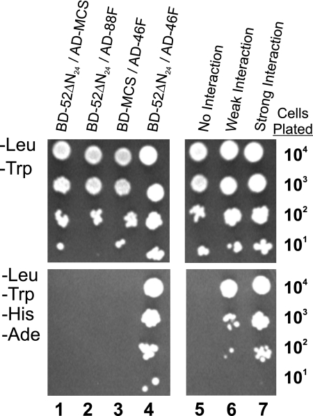
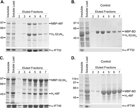
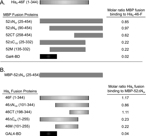
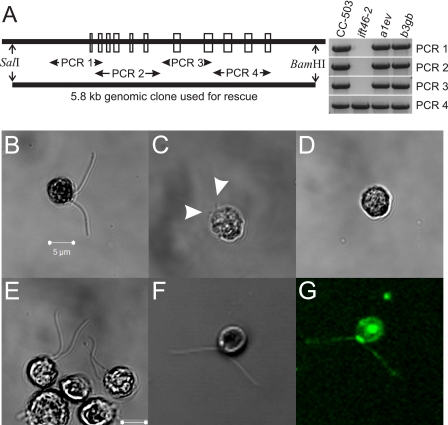
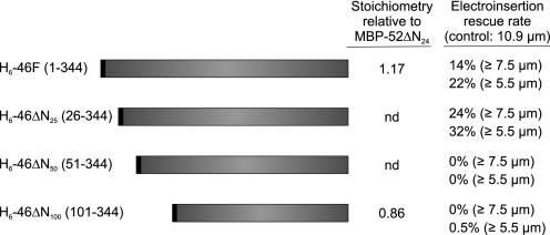
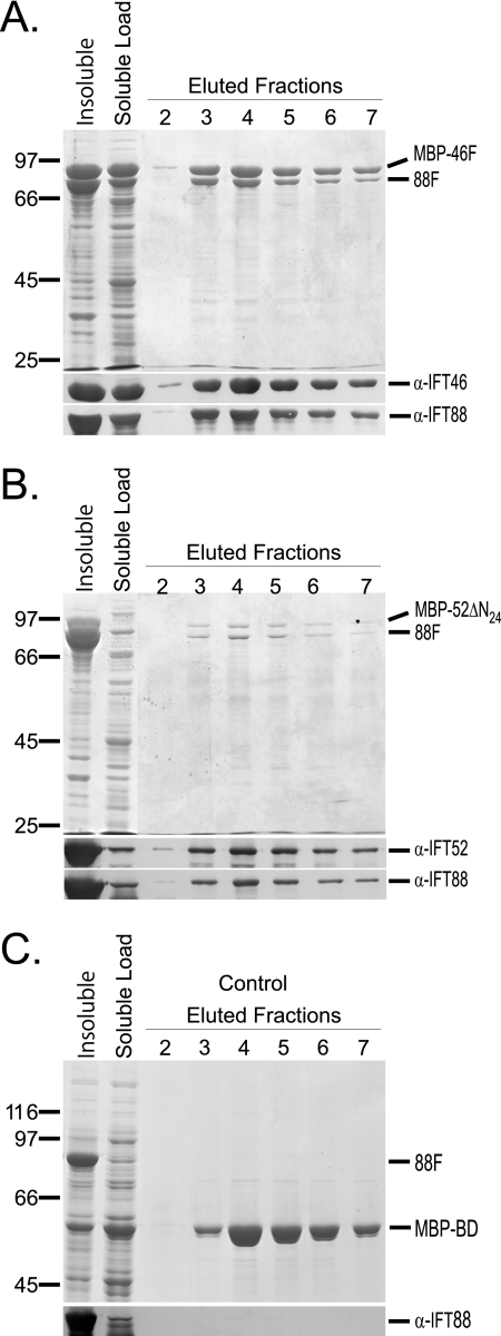
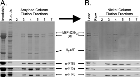
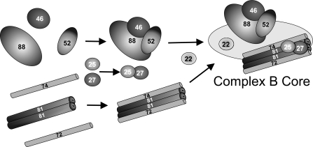
Intraflagellar transport (IFT) particles of Chlamydomonas reinhardtii contain two distinct protein complexes, A and B, composed of at least 6 and 15 protein subunits, respectively. As isolated from C. reinhardtii flagella, IFT complex B can be further reduced to a approximately 500-kDa core that contains IFT88, 2x IFT81, 2x IFT74/72, IFT52, IFT46, IFT27, IFT25, and IFT22. In this study, yeast-based two-hybrid analysis was combined with bacterial coexpression to show that three of the core B subunits, IFT88, IFT52, and IFT46, interact directly with each other and, together, are capable of forming a ternary complex. Chemical cross-linking results support the IFT52-IFT88 interaction and provide additional evidence of an association between IFT27 and IFT81. With previous studies showing that IFT81 and IFT74/72 interact to form a (IFT81)(2)(IFT74/72)(2) heterotetramer and that IFT27 and IFT25 form a heterodimer, the architecture of complex B is revealing itself. Last, electroporation of recombinant IFT46 was used to rescue flagellar assembly of a newly identified ift46 mutant and to monitor in vivo localization and movement of the IFT particles.


Figures

FIGURE 1.
FIGURE 1.
Direct interaction of IFT46 and IFT52 using yeast-based two-hybrid analysis. Serial dilutions of YRG-2 yeast containing both AD and BD plasmid constructs were grown on selective (−Leu, −Trp) medium (top panels) to verify the presence of both AD and BD plasmids and a more restrictive (−Leu, −Trp, −His, −Ade) medium (bottom panels) to test for protein interactions. Column 1, the lack of growth in the absence of histidine and adenine revealed that IFT52 did not interact with the control AD protein. Column 2, the lack of cell growth on the more restrictive medium (−His, −Ade) revealed that IFT52 and IFT88 failed to interact in this assay. Column 3, IFT46 (AD-46F) showed no interaction with the control BD protein. Column 4, the combination of the BD-52ΔN24 and AD-46F plasmids conferred growth in the absence of histidine and adenine, which was consistent with a direct interaction between IFT52 and IFT46. Columns 5–7, negative, weak, and strong interaction control plasmids are described under “Experimental Procedures.”
FIGURE 2.
FIGURE 2.
Coexpression and copurification of recombinant IFT46 and IFT52 using amylose affinity chromatography. The first two lanes of each Coomassie Blue-stained gel contain insoluble and soluble fractions of bacterial cell lysates. The soluble fraction was loaded onto amylose MBP affinity resin, washed with column buffer (ACB), and eluted using 10 mm maltose in ACB; fractions 2–7 are shown here. Western blots were probed with either anti-IFT52 or anti-IFT46, as indicated. A, following coexpression, H6-52ΔN24 coeluted with MBP-46F. B, H6-52ΔN24 did not copurify with the MBP-BD control. C, following coexpression, H6-46F coeluted with MBP-52ΔN24. D, H6-46F did not copurify with the MBP-BD control.
FIGURE 3.
FIGURE 3.
Interactions between IFT46 and IFT52 are mediated by C-terminal domains. Pairwise combinations of proteins were coexpressed. Interactions were monitored using MBP affinity chromatography. Molar ratios of copurified proteins were determined using densitometric scanning of Coomassie Blue-stained gels. A, full-length IFT46 (His6-46F) was coexpressed with various MBP-tagged IFT52 deletion and control proteins. B, nearly full-length IFT52 (MBP-52ΔN24) was coexpressed with various His6-46 deletion and control proteins.
FIGURE 4.
FIGURE 4.
Electroporation of recombinant His6-46F rescues the ift46-2 flagellar assembly phenotype. A, a screen of random insertional motility mutants revealed a C. reinhardtii strain, ift46-2, that carries a deletion of most of the IFT46 gene; the exons of the wild-type IFT46 are depicted by open boxes. The lack of specific PCR amplification products (PCR 1–3) revealed that most of the IFT46 gene was disrupted in the ift46-2 strain. A 5809 bp SalI/BamHI genomic fragment was used to rescue the ift46-2 flagellar assembly phenotype. PCR analysis of two rescued strains, a1ev and b3gb, revealed that the IFT46 gene had been successfully reintroduced. B, the cell wall-deficient parental CC-503 strain assembles flagella of normal length and function. C, although most ift46-2 cells were bald, ∼6% were able to assemble short flagella (average ∼3 μm) as indicated by arrowheads. D, electroporation of BSA into ift46-2 resulted in no change in the bald phenotype; the image shown was taken 4 h postelectroporation. E, at 4 h postelectroporation of recombinant His6-IFT46, many cells displayed partial or full assembly of motile flagella. F and G, electroporation of Alexa-fluor 488-labeled His6-46F resulted in similar rates of rescue; 4-h postelectroporation images shown are white light (F) and emission at 518 nm (G). Many flagellated cells displayed concentrated pools of fluorescent IFT46 near the basal bodies with punctate staining throughout the flagella. Intraflagellar transport of the labeled IFT46 could also be observed in some cells.
FIGURE 5.
FIGURE 5.
The N-terminal 25 amino acids of IFT46 are not required for recombinant protein rescue. Recombinant His6-46 proteins with the indicated N-terminal deletions were purified by metal chelate chromatography and introduced into ift46-2 cells using electroporation with a final concentration of recombinant protein at 0.1 mg/ml. For comparison, the stoichiometry of the full-length IFT46 (H6-46F) and the N-terminal 100-amino acid deletion (H6-46ΔN100) relative to the MBP-tagged IFT52 are shown (see Fig. 3). Both the full-length and N-terminal 25-amino acid deletion IFT46 proteins were capable of similar rates of flagellar assembly rescue 8 h postelectroporation. nd, not determined.
FIGURE 6.
FIGURE 6.
IFT88 interacts separately with either IFT52 or IFT46. Untagged IFT88 was coexpressed separately with MBP fusions of IFT52, IFT46, and BD (negative control) proteins. The first two lanes of each gel contain the insoluble and soluble fractions of bacterial cell lysates. The soluble fraction was loaded onto amylose MBP-affinity resin and washed with ACB buffer prior to 10 mm maltose elution. Elution fractions 2–7 are shown on the Coomassie Blue-stained SDS-polyacrylamide gels. Corresponding Western blots are shown below each gel. A and B, the untagged IFT88 copurifies with MBP-46F and MBP-52ΔN24, respectively. C, IFT88 does not copurify with the MBP-BD control protein.
FIGURE 7.
FIGURE 7.
Copurification of H6-46F, MBP-52ΔN24, and IFT88 using tandem affinity chromatography. Upper panels, Coomassie Blue-stained SDS-polyacrylamide gels; lower panels, transfer membranes probed with antibodies directed against IFT88, IFT52, or IFT46. A, MBP affinity purification of coexpressed recombinant H6-46F, MBP-52ΔN24, and untagged IFT88. Soluble bacterial lysate was loaded onto an amylose column. After washing the resin, MBP-52ΔN24 and associated proteins were eluted using 10 mm maltose; fractions 2–7 are shown here. Both H6-46F and the untagged IFT88 coeluted with the MBP-52ΔN24. B, the peak fractions from the MBP affinity chromatography (A) were pooled and further purified using metal (Ni2+) chelate chromatography. Elution of the H6-46F from the Ni2+ resin using imidazole resulted in the coelution of both MBP-52ΔN24 and IFT88, indicating that the three proteins were capable of forming a stable ternary complex.
FIGURE 8.
FIGURE 8.
Hypothetical model of in vivo assembly of the IFT complex B core. IFT46, IFT52, and IFT88 form a ternary complex prior to assembly with the IFT81, IFT74/72 tetramer. Previous studies have shown that the IFT81 and IFT74/72 subunits and the IFT27 and IFT25 subunits are capable of forming stable associations in the absence of other complex B proteins (33). The actual order in which these and additional subunits assemble onto the core is unknown.

Similar articles

Cited by

References

    1. Wheatley D. N. (1995) Pathobiology 63, 222–238 - PubMed
    1. Bray D. (2001) Cell Movements: From Molecules to Motility, pp. 3–16, 225–241, Garland Publishing, New York, NY
    1. Praetorius H. A., Spring K. R. (2005) Annu. Rev. Physiol. 67, 515–529 - PubMed
    1. Davis E. E., Brueckner M., Katsanis N. (2006) Dev. Cell 11, 9–19 - PubMed
    1. Pazour G. J., Witman G. B. (2003) Curr. Opin. Cell Biol. 15, 105–110 - PubMed

Publication types