. 2010 Apr;22(4):1344-57.
doi: 10.1105/tpc.109.072637. Epub 2010 Apr 30.

Endocytic and secretory traffic in Arabidopsis merge in the trans-Golgi network/early endosome, an independent and highly dynamic organelle

Affiliations

Endocytic and secretory traffic in Arabidopsis merge in the trans-Golgi network/early endosome, an independent and highly dynamic organelle

Corrado Viotti et al. Plant Cell. 2010 Apr.

Abstract

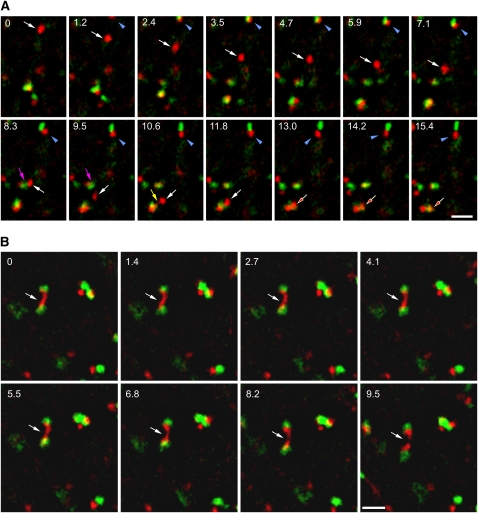
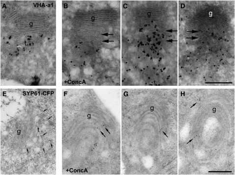
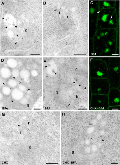
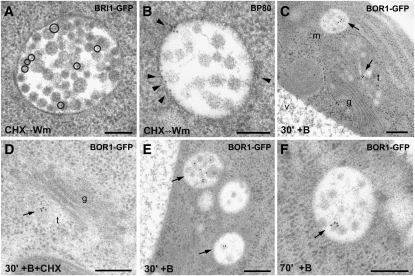
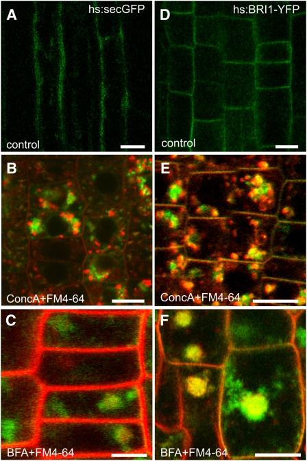
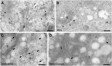
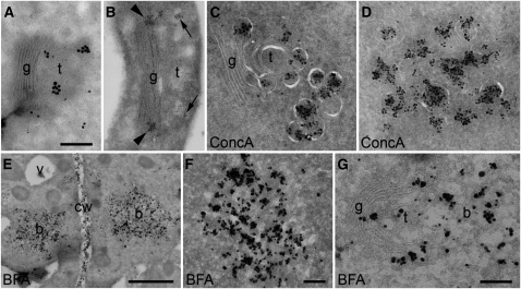
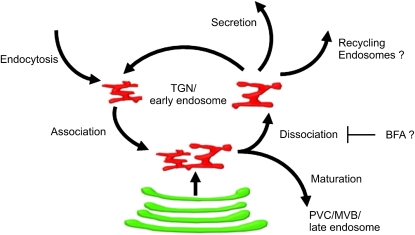
Plants constantly adjust their repertoire of plasma membrane proteins that mediates transduction of environmental and developmental signals as well as transport of ions, nutrients, and hormones. The importance of regulated secretory and endocytic trafficking is becoming increasingly clear; however, our knowledge of the compartments and molecular machinery involved is still fragmentary. We used immunogold electron microscopy and confocal laser scanning microscopy to trace the route of cargo molecules, including the BRASSINOSTEROID INSENSITIVE1 receptor and the REQUIRES HIGH BORON1 boron exporter, throughout the plant endomembrane system. Our results provide evidence that both endocytic and secretory cargo pass through the trans-Golgi network/early endosome (TGN/EE) and demonstrate that cargo in late endosomes/multivesicular bodies is destined for vacuolar degradation. Moreover, using spinning disc microscopy, we show that TGN/EEs move independently and are only transiently associated with an individual Golgi stack.


Figures

Figure 1.
Figure 1.
The TGN Is an Independent Organelle That Undergoes Reversible Homotypic Association. Spinning disc confocal microscopy of hypocotyl cells expressing VHA-a1-RFP and ST-GFP. Numbers = seconds; bars = 5 μM. (A) The TGN is a highly mobile and independent organelle (white arrows, time point from 0 to 11.8 s) that temporarily pauses and becomes closely associated with a Golgi stack (blue arrowheads, 1.2 to 15.4 s). Homotypic association of individual TGNs was also observed (red arrows, 13.0 to 15.4 s), and sometimes a thin protrusion between the interacting partners was visible (yellow arrows, 10.6 s). Not all TGNs are competent for this interaction (magenta arrow, 8.3 and 9.5 s). (B) A dissociation event for an elongated TGN shared by two Golgi stacks (white arrows, 0 to 9.5 s).
Figure 2.
Figure 2.
The Identity and Independence of the TGN Is Affected by ConcA. Immunogold labeling of VHA-a1 and SYP61-CFP in ConcA-treated cells. (A) to (D), silver-enhanced cryosections; (E) to (H), HM20 resin sections (see Methods). Bars = 250 nm. (A) and (E) VHA-a1 and SYP61 are highly specific markers for the plant TGN/EE. (B) to (D) and (F) to (H) In the presence of ConcA, VHA-a1 and SYP61-CFP were additionally found on Golgi stacks, and a clear spatial separation between the Golgi apparatus and the TGN was no longer recognizable. Furthermore, ConcA induces the proliferation of the Golgi cisternae, accompanied by a loss of identity of the two compartments.
Figure 3.
Figure 3.
Endocytosed BRI1-GFP Is Found at the TGN and in MVBs. Immunogold labeling and CLSM analyses of root tips from BRI1-GFP Arabidopsis plants. m, multivesicular body; t, TGN; g, Golgi; er, endoplasmic reticulum; b, BFA compartment. The symbol “→” means that the second inhibitor was added with the first still present. IEM bars = 200 nm; CLSM bars = 5 μm. (A) BRI1-GFP localizes to both the TGN and MVB (arrowheads). (B) The TGN is positively labeled (arrowheads) also when it is not closely associated with the Golgi stacks. (C) The core of the BFA compartment is labeled, and a surrounding punctate pattern is also visible (arrows). (D) and (E) IEM confirmed the localization of BRI1-GFP in the core of the BFA compartment and on the MVB (arrowheads). (F) The protein synthesis inhibitor CHX does not prevent the accumulation of BRI1-GFP in the core of the BFA compartment (arrowheads). (G) In the presence of CHX, BRI1 is still detected at the TGN (arrowheads). (H) In the presence of CHX and BFA, BRI1 is detected in the BFA compartment (arrowheads).
Figure 4.
Figure 4.
BRI1-GFP and BOR1-GFP, but Not BP80, Are Found on the Inner Vesicles of MVBs. Immunogold labeling of BP80, BRI1-GFP, or BOR1-GFP in Arabidopsis root tips. m, multivesicular body; t, TGN; v, vacuole; g, Golgi. Bars = 200 nm. (A) BRI1-GFP is found on the inner vesicles of MVBs (highlighted by circles). (B) By contrast, BP80 localizes on the boundary membrane of MVBs (arrowheads). (C) BOR1-GFP localizes both to the TGN and to the MVB after 30 min of exposure to boron (arrowheads). (D) BOR1-GFP localizes to the TGN (arrow) after 30 min of exposure to boron also in the presence of CHX. (E) and (F) BOR1-GFP always localizes to the inner vesicles of MVBs, after either 30 or 70 min of exposure to boron.
Figure 5.
Figure 5.
The Effects of ConcA and BFA on hs:secGFP and hs:BRI1-YFP. CLSM images of cells in the root elongation zone of hs:secGFP ([A] to [C]) or hs:BRI1-YFP ([D] to [F]) seedlings. Bars = 10 μM. (A) and (D) Untreated cells expressing secGFP (A) or BRI1-YFP (D) 5 h after heat shock. (B) and (E) FM4-64 was added 5 min before ConcA. Thirty minutes after addition of ConcA, the heat shock started. After 5 h of expression, a strong intracellular signal of both secGFP (B) or BRI1-YFP (E) was detectable. (C) and (F) BFA was added 30 min before the heat shock. Five hours after the heat shock, root cells were stained with FM4-64 for 30 min. Both secGFP (C) and BRI1-YFP (F) were detected in the core of the BFA compartment.
Figure 6.
Figure 6.
Newly Synthesized BRI1-YFP Passes through Both the Golgi and the TGN. Immunogold labeling of hs:BRI1-YFP in Arabidopsis root tips in the presence of different combinations of inhibitors. g, Golgi; t, TGN; m, MVB; b, BFA compartment. Bars = 200 nm. (A) After a 1-h heat shock treatment, seedlings were immediately high-pressure frozen and freeze substituted. hs:BRI1-YFP localizes both to the Golgi apparatus and the TGN (arrows). (B) Treatment with the endocytosis inhibitor Wm started 30 min before the heat shock. hs:BRI1-YFP again localized to both the Golgi and the TGN (arrows), whereas the MVB was not labeled. (C) Treatment with Wm and BFA started 30 min before the heat shock. hs:BRI1-YFP localized both to the Golgi apparatus and the core of the BFA compartment (arrows). (D) Treatment with Wm and ConcA started 30 min before the heat shock. hs:BRI1-YFP localized to the TGN (arrows).
Figure 7.
Figure 7.
Xyloglucans Accumulate after ConcA Treatment and in BFA Compartments. Immunogold labeling of a fucosylated xyloglucan epitope with CCRC-M1 antibodies. b, BFA compartment; cw, cell wall; g, Golgi stack; t, TGN. Bars = 250 nm in (A) to (D), (F), and (G) and 2 μm in (E). (A) Labeling of the rims of Golgi stacks and TGN with silver-enhanced Nanogold-F(ab)2. (B) Labeling of the rims of Golgi stacks (arrowheads) and TGN (arrows) with 12-nm gold particles coupled to IgG. Clustered marker molecules represent secretory vesicles. (C) and (D) Labeling of aggregated large Golgi-derived secretory vesicles after treatment with ConcA. (C) Vesicles close to a Golgi stack. (E) to (G) Labeling of BFA compartments after treatment with BFA. (E) Overview showing two vesicles aggregates. (F) Enlarged BFA compartment. (G) BFA compartment with attached Golgi stack.
Figure 8.
Figure 8.
Model Illustrating the Multiple Functions of the TGN in Plants. The TGN can be found either separated or closely associated to the Golgi stack. TGNs can associate homotypically, and the process is reversible. BFA causes the formation of large TGN-derived agglomerates (BFA compartments); we propose that this drug inhibits the process of dissociation between TGNs.

References

    1. An Q., van Bel A.J., Huckelhoven R. (2007). Do plant cells secrete exosomes derived from multivesicular bodies? Plant Signal. Behav. 2: 4–7 - PMC - PubMed
    1. Baluska F., Hlavacka A., Samaj J., Palme K., Robinson D.G., Matoh T., McCurdy D.W., Menzel D., Volkmann D. (2002). F-actin-dependent endocytosis of cell wall pectins in meristematic root cells. Insights from brefeldin A-induced compartments. Plant Physiol. 130: 422–431 - PMC - PubMed
    1. Baluska F., Liners F., Hlavacka A., Schlicht M., Van Cutsem P., McCurdy D.W., Menzel D. (2005). Cell wall pectins and xyloglucans are internalized into dividing root cells and accumulate within cell plates during cytokinesis. Protoplasma 225: 141–155 - PubMed
    1. Batoko H., Zheng H.Q., Hawes C., Moore I. (2000). A rab1 GTPase is required for transport between the endoplasmic reticulum and Golgi apparatus and for normal Golgi movement in plants. Plant Cell 12: 2201–2218 - PMC - PubMed
    1. Bonifacino J.S., Hurley J.H. (2008). Retromer. Curr. Opin. Cell Biol. 20: 427–436 - PMC - PubMed

Publication types

MeSH terms

LinkOut - more resources