. 2010 May 13;465(7295):243-7.
doi: 10.1038/nature08966. Epub 2010 May 2.

Histone H2A deubiquitinase activity of the Polycomb repressive complex PR-DUB

Affiliations

Histone H2A deubiquitinase activity of the Polycomb repressive complex PR-DUB

Johanna C Scheuermann et al. Nature. .

Abstract

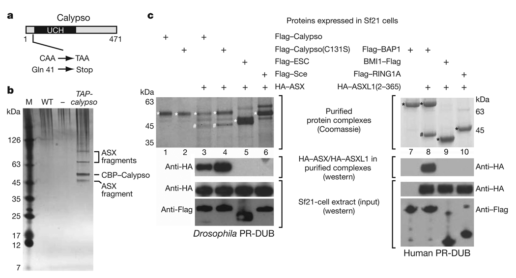
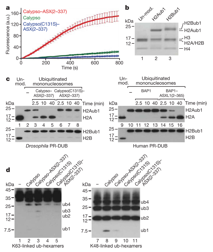
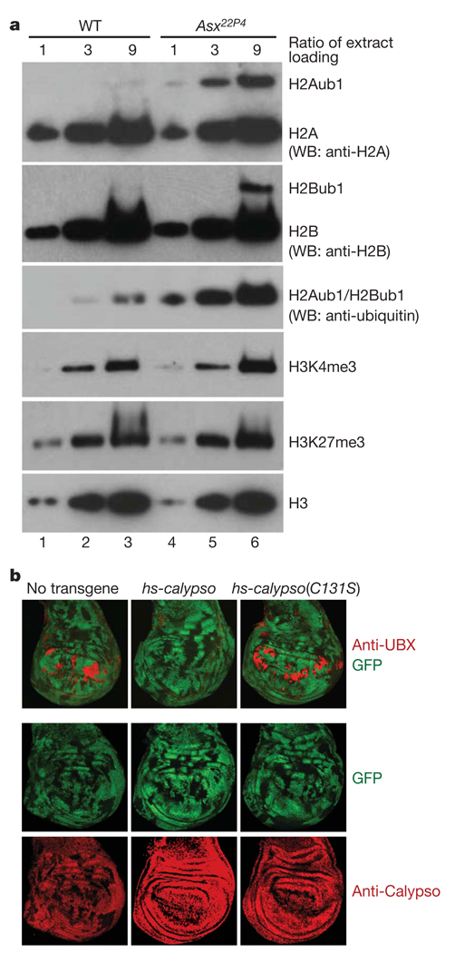
Polycomb group (PcG) proteins are transcriptional repressors that control processes ranging from the maintenance of cell fate decisions and stem cell pluripotency in animals to the control of flowering time in plants. In Drosophila, genetic studies identified more than 15 different PcG proteins that are required to repress homeotic (HOX) and other developmental regulator genes in cells where they must stay inactive. Biochemical analyses established that these PcG proteins exist in distinct multiprotein complexes that bind to and modify chromatin of target genes. Among those, Polycomb repressive complex 1 (PRC1) and the related dRing-associated factors (dRAF) complex contain an E3 ligase activity for monoubiquitination of histone H2A (refs 1-4). Here we show that the uncharacterized Drosophila PcG gene calypso encodes the ubiquitin carboxy-terminal hydrolase BAP1. Biochemically purified Calypso exists in a complex with the PcG protein ASX, and this complex, named Polycomb repressive deubiquitinase (PR-DUB), is bound at PcG target genes in Drosophila. Reconstituted recombinant Drosophila and human PR-DUB complexes remove monoubiquitin from H2A but not from H2B in nucleosomes. Drosophila mutants lacking PR-DUB show a strong increase in the levels of monoubiquitinated H2A. A mutation that disrupts the catalytic activity of Calypso, or absence of the ASX subunit abolishes H2A deubiquitination in vitro and HOX gene repression in vivo. Polycomb gene silencing may thus entail a dynamic balance between H2A ubiquitination by PRC1 and dRAF, and H2A deubiquitination by PR-DUB.


Figures

Figure 1
Figure 1. The Polycomb group proteins BAP1 and ASX form a conserved complex in vivo and in vitro
a, Domain architecture of the Drosophila Calypso protein and molecular lesions in calypso mutant alleles. UCH, ubiquitin C-terminal hydrolase domain. b, Calypso complexes isolated by TAP from wild-type (WT) or TAP-calypso transgenic embryos. Input material for purification was normalized by protein concentration, and equivalent amounts of eluate from calmodulin-affinity resin were separated on a 4–12% polyacrylamide gel and visualized by silver staining together with a molecular mass marker (M). Calypso bait protein containing the calmodulin-binding tag (CBP–Calypso), and bands representing ASX fragments were identified by mass spectrometry (Supplementary Table 1 and Supplementary Fig. 2). No band corresponding to full-length ASX (180 kDa) was detected in several independent purifications, even though ASX is present as a single polypeptide of 180 kDa in total embryo extracts (Supplementary Fig. 5). This suggests that ASX is degraded during nuclear extract preparation or TAP purification. c, Reconstitution of recombinant Calypso–ASX and BAP1–ASXL1 complexes. Proteins were extracted by Flag-affinity purification from cell lysates containing the indicated Flag-tagged proteins and HA–ASX(2–337) (left) or HA–ASXL1(2–365) (right). Experiments with full-length ASX are shown in Supplementary Fig. 3. Proteins were visualized by Coomassie staining or western blotting analysis, as indicated. Input material for experiments in lanes 3–6 (left) and 7–10 (right) were probed by western blotting to ensure that comparable amounts of proteins were present in cell lysates. On the Coomassie-stained gel, Flag-tagged proteins are marked with an asterisk, HA–ASX(2–337) and HA–ASXL1(2–365) are marked with a hash symbol.
Figure 2
Figure 2. PR-DUB is bound at Polycomb target genes in Drosophila
a, PR-DUB is bound at PREs of PcG target genes in Drosophila. ChIP profiles of PR-DUB subunits ASX (dark blue) and Calypso (light blue), and of Ph (grey) and Pho (grey) at the Antennapedia HOX gene cluster in imaginal disc and CNS tissues from third instar Drosophila larvae. Hybridization intensities for oligonucleotide probes are plotted as coloured bars above the genomic map (release 5, kilobase coordinates) of Drosophila melanogaster; significantly enriched regions are marked below plots. HOX genes labial (lab), proboscipedia (pb), Deformed (Dfd), Sex combs reduced (Scr), Antennapedia (Antp) and other genes on the plus (above) or minus (below) strand are represented with exons (black boxes) and introns (thin black lines). b, Venn diagrams showing the overlap of 879 PR-DUB-bound regions with 1,681 Ph-bound and 670 Pho-bound regions in larval cells. c, PR-DUB is bound at the inactive and at the active Ubx gene. ChIP analyses monitoring ASX and Calypso binding in wing and haltere/third leg imaginal discs from wild-type third instar Drosophila larvae. Graphs show results from independent ChIP reactions (n = 3 ChIP reactions) with ASX or Calypso antibodies. ChIP signals, measured by qPCR, are presented as the mean percentage of input chromatin precipitated at each region; error bars indicate±s.d. (see Methods). Locations of Ubx PREs (boxes) and other regions relative to the Ubx transcription start site are indicated in kilobases. As control, binding was monitored at two euchromatic (eu) and one heterochromatic (het) region elsewhere in the genome, and at the PREs of the HOX genes Abd-B and Scr that are both inactive in wing and haltere/third leg imaginal discs. Calypso and ASX are bound at Ubx PREs both in wing and in haltere/third leg disc cells; at the −30-kb PRE, Calypso and ASX ChIP signals were comparable in wing and haltere/third leg chromatin, at the kb PRE, the signal in haltere/third leg chromatin is about 2–4-fold lower than in wing chromatin, paralleling PRC1 and PRC2 binding at both these PREs.
Figure 3
Figure 3. Recombinant Drosophila and human PR-DUB deubiquitinate H2A in nucleosomes in vitro
a, Cleavage of Ub-AMC by Calypso and Calypso–ASX(2–337) complexes. Reactions (n = 4) contained 25 pmol Ub-AMC and 10 pmol of the indicated protein (complex); release of AMC was monitored by fluorescence spectroscopy at 436 nm; error bars indicate ±s.d. a.u., arbitrary units. b, Mononucleosomes were reconstituted with recombinant Xenopus histone octamers and were unmodified (lane 1), monoubiquitinated at H2AK119 (lane 2) (see Methods) or monoubiquitinated at H2BK120 (lane 3) (see Methods). The material was analysed on a 4–12% polyacrylamide gradient gel and histones were visualized by Coomassie staining. c, Drosophila and human PR-DUB deubiquitinate H2Aub1 in nucleosomes. Xenopus mononucleosomes (15 pmol) containing 30 pmol of either H2Aub1 (top gels left and right) or H2Bub1 (bottom gels left and right) were incubated without (lanes 2 and 10) or with 30 pmol of the indicated Drosophila PR-DUB complexes (lanes 3–8) or human BAP1 or PR-DUB complex (lanes 11–16), respectively, and deubiquitination was monitored at indicated time points by western blot analysis with anti-H2A (top gels left and right) or anti-H2B (bottom gels left and right) antibody (5 pmol nucleosome per lane). Unmodified mononucleosomes (lanes 1 and 9) served as a control. Comparable results were obtained with Drosophila mononucleosomes containing H2Aub1 (Supplementary Fig. 4). PR-DUB containing full-length ASX(1–1668) also specifically deubiquitinated H2Aub1 but not H2Bub1 in nucleosomes (Supplementary Fig. 4). d, K48- or K63-linked hexameric polyubiquitin chains (160 ng; corresponding to maximally 17.5 pmol ubiquitin linkage bonds) were incubated for 40 min with 10 pmol of the indicated protein or protein complex under the same assay conditions as in c, followed by western blot analysis with an anti-ubiquitin antibody.
Figure 4
Figure 4. PR-DUB is required for H2A deubiquitination in Drosophila and its catalytic activity is essential for HOX gene repression
a, PR-DUB is required for H2A deubiquitination in Drosophila embryos. Serial dilutions (1:3:9) of histone extracts from 16–18-h-old wild-type or Asx22P4 homozygous embryos were separated on 4–12% polyacrylamide gels and analysed by western blotting with the indicated antibodies. H2Aub1 levels in lanes 3 and 4 are comparable, suggesting that H2Aub1 levels are almost tenfold higher in Asx22P4 mutants than in wild type. H2Bub1 levels in Asx22P4 mutants are less than threefold increased compared to wild type (compare lane 3 with lanes 5 and 6). The band detected by an anti-ubiquitin antibody represents the combined signal of H2Aub1 and H2Bub1. H3K4me3 levels appear very slightly increased in Asx22P4 mutants. b, Calypso deubiquitinase activity is required for HOX gene repression. Wing imaginal discs with clones of calypso2 homozygous mutant cells from animals that carried no transgene or the indicated hsp70-calypso transgenes. calypso2 mutant cells are marked by the absence of GFP and discs were stained with antibodies against UBX or Calypso protein, as indicated. In all cases, clones were induced 96 h before analysis and larvae were repeatedly heat-shocked for 1 h every 12 h over a 96-h period to provide a continuous supply of Calypso protein from the transgene. In the absence of an hsp70-calypso transgene, Ubx is misexpressed in most calypso2 mutant clones in the pouch of the disc but remains repressed in the notum and hinge (left). Wild-type Calypso protein rescues repression of Ubx in mutant clones (middle), whereas the Calypso(C131S) protein fails to rescue (right), even though both transgene-encoded proteins are expressed at comparable levels and show nuclear localization like endogenous Calypso protein (bottom row).

Comment in

  • The DUBle life of polycomb complexes.
    Schuettengruber B, Cavalli G. Schuettengruber B, et al. Dev Cell. 2010 Jun 15;18(6):878-80. doi: 10.1016/j.devcel.2010.06.001. Dev Cell. 2010. PMID: 20627069

References

    1. Schwartz YB, Pirrotta V. Polycomb silencing mechanisms and the management of genomic programmes. Nature Rev. Genet. 2007;8:9–22. - PubMed
    1. Schuettengruber B, Chourrout D, Vervoort M, Leblanc B, Cavalli G. Genome regulation by polycomb and trithorax proteins. Cell. 2007;128:735–745. - PubMed
    1. Müller J, Verrijzer CP. Biochemical mechanisms of gene regulation by polycomb group protein complexes. Curr. Opin. Genet. Dev. 2009;19:150–158. - PubMed
    1. Simon JA, Kingston RE. Mechanisms of polycomb gene silencing: knowns and unknowns. Nature Rev. Mol. Cell Biol. 2009;10:697–708. - PubMed
    1. Pietersen AM, van Lohuizen M. Stem cell regulation by polycomb repressors: postponing commitment. Curr. Opin. Cell Biol. 2008;20:201–207. - PubMed

Publication types

MeSH terms