. 2010 Jun;6(6):433-41.
doi: 10.1038/nchembio.367. Epub 2010 May 2.

Cyclooxygenase-2 generates anti-inflammatory mediators from omega-3 fatty acids

Affiliations

Cyclooxygenase-2 generates anti-inflammatory mediators from omega-3 fatty acids

Alison L Groeger et al. Nat Chem Biol. 2010 Jun.

Abstract

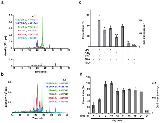
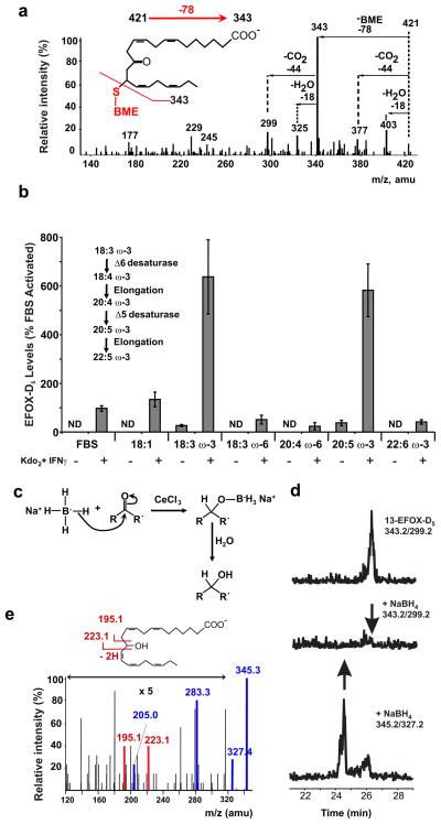
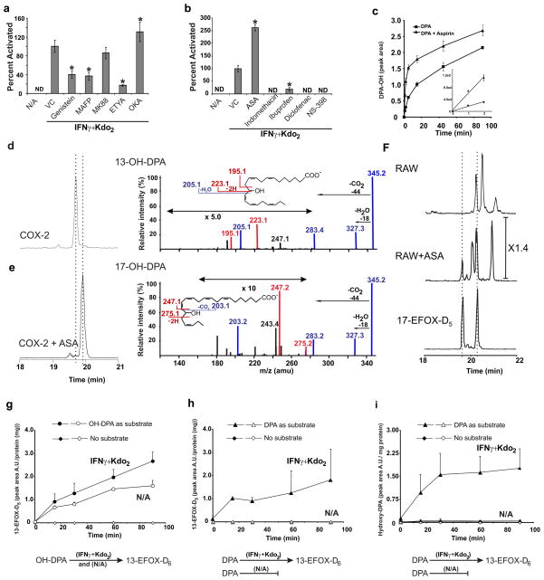
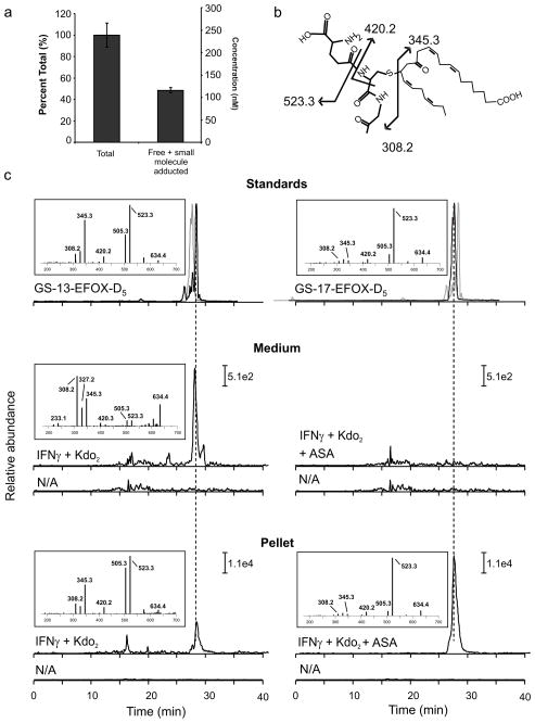
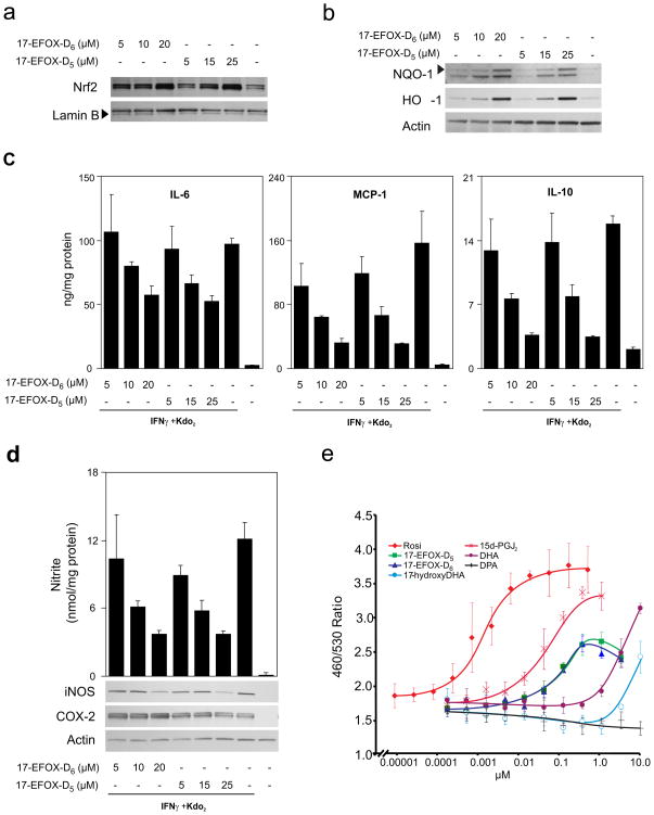
Electrophilic fatty acids are generated during inflammation by non-enzymatic reactions and can modulate inflammatory responses. We used a new mass spectrometry-based electrophile capture strategy to reveal the formation of electrophilic oxo-derivatives (EFOX) from the omega-3 fatty acids docosahexaenoic acid (DHA) and docosapentaenoic acid (DPA). These EFOX were generated by a cyclooxygenase-2 (COX-2)-catalyzed mechanism in activated macrophages. Modulation of COX-2 activity by aspirin increased the rate of EFOX production and their intracellular levels. Owing to their electrophilic nature, EFOX adducted to cysteine and histidine residues of proteins and activated Nrf2-dependent anti-oxidant gene expression. We confirmed the anti-inflammatory nature of DHA- and DPA-derived EFOX by showing that they can act as peroxisome proliferator-activated receptor-gamma (PPAR gamma) agonists and inhibit pro-inflammatory cytokine and nitric oxide production, all within biological concentration ranges. These data support the idea that EFOX are signaling mediators that transduce the beneficial clinical effects of omega-3 fatty acids, COX-2 and aspirin.


Conflict of interest statement

Conflict of Interest: BAF acknowledges financial interest in Complexa, Inc.

Figures

Figure 1
Figure 1. EFOXs are produced during macrophage activation
a, MRM scans following the neutral loss of 78 reveal EFOXs adducted with BME in cell extracts from activated (upper) and non-activated (N/A, lower) RAW 264.7 cells. b, EFOX levels in PMA-differentiated THP-1 cells 8 h post activation. c, EFOX-D5 levels in RAW264.7 cells activated with the indicated compounds at the following concentrations: LPS (0.5 μg/ml), Kdo2 (0.5 μg/ml), IFNγ (200 U/ml), PMA (3.24 μM), fMLP (1 μM). Data are expressed as mean ± S.D. (n=4), where * = significantly different (p<0.01) from “PMA + IFNγ + LPS,” and # = significant difference (p<0.01) between “LPS” and “Kdo2 + IFNγ” (one-way ANOVA, post-hoc Tukey’s test). d, EFOX-D5 levels in RAW264.7 cells were quantified at the indicated time points post activation. ND indicates not detectable.
Figure 2
Figure 2. EFOX-D5 is an α,β-unsaturated oxo-derivative of DPA
a, Characteristic BME-EFOX fragmentation pattern derived from the EPI of BME-EFOX-D5. b, EFOX-D5 levels in activated RAW264.7 cells grown for 3 days in medium supplemented with the indicated FA. ND indicates not detectable. c, Diagram of NaBH4 reaction. d, MRM scans monitoring for the m/z transitions 343.2/299.2 (13-EFOX-D5/loss of CO2; upper and middle panel) and 345.2/327.2 (hydroxy-DPA/loss of H2O; lower panel) in RAW264.7 cell lysates purified for EFOX-D5 ± NaBH4. e, MS/MS fragmentation of EFOX-D5 purified from activated RAW 264.7 cells and reduced with NaBH4.
Figure 3
Figure 3. EFOX-D5 formation is dependent on COX-2 activity
a, b, EFOX-D5 levels in RAW264.7 activated and treated with the indicated inhibitors at the following concentrations: genistein (25 μM), MAFP (25 μM), MK886 (500 nM), ETYA (25 μM), OKA (50 nM), ASA (200 μM), indomethacin (25 μM), ibuprofen (100 μM), diclofenac (1 μM) and NS-398 (4 μM). ND indicates not detectable. Data are expressed as mean ± S.D. (n=4), where * = significantly different (p<0.01) from “Kdo2 + IFNγ” (one-way ANOVA, post-hoc Tukey’s test). c, Temporal formation of hydroxy-precursors (MRM 345/327) of EFOX-D5 synthesized in vitro by purified ovine COX-2 + DPA ± ASA. d, e, Chromatographic profiles (left panels) and spectra (right panels) of the two isomers formed by COX-2 ± ASA. f, BME-adducted 17-EFOX-D5 standard (MRM 421.2/343.2) and EFOX-D5 BME-adducts from activated RAW264.7 cells ± ASA. g–i, Production of EFOX-D5 and OH-DPA by RAW264.7 cell lysates supplemented with OH-DPA, DPA, or vehicle alone. Full and empty symbols indicate activated and non-activated cell lysates respectively.
Figure 4
Figure 4. EFOXs form adducts with proteins and GSH following activation of RAW264.7
a, Cell lysates from activated RAW264.7 cells were split into two groups: treatment with BME followed by protein precipitation with acetonitrile (“Total”) and protein precipitation followed by BME treatment (“Free + small molecule adducted”). b, Chemical structure and fragmentation pattern of GS-13-EFOX-D5. c, Chromatographic profiles and positive ion mass spectra of GSH adducts of 13-EFOX-D5 and 17-EFOX-D5 derived from standards (upper panels), cell medium (middle panel) and cell pellet (lower panel), N/A profiles correspond to non-activated cell samples. Grey chromatograms represent GS-17-EFOX-D5 (upper panel left) and GS-13-EFOX-D5 (upper panel right) standards. Fragments 345.3 and 523.3 were monitored in cell media and cell pellet samples respectively.
Figure 5
Figure 5. 17-EFOX-D6 and 17-EFOX-D5 modulate anti-oxidant and inflammatory responses
a, b, RAW264.7 cells treated with 17-EFOX-D6 and 17-EFOX-D5 were harvested at 1 h for nuclear Nrf2 (a) and 18 h for of HO-1 and NQO-1 (b). c, d, RAW264.7 cells pretreated for 6 h with 17-EFOX-D6 and 17-EFOX-D5 were harvested 12 h post activation and IL-6, MCP-1, IL-10 levels, and nitrite levels were measured in the cell media and normalized to protein. d, iNOS and COX-2 levels were detected in cell lysates. e, PPARγ beta-lactamase reporter assays for Rosiglitazone, 17-EFOX-D6, 17-EFOX-D5, 15d-PGJ2, 17-hydroxyDHA, DPA, and DHA.

Comment in

References

    1. Connor WE. Importance of n-3 fatty acids in health and disease. Am J Clin Nutr. 2000;71:171S–175S. - PubMed
    1. Neuringer M, Anderson GJ, Connor WE. The essentiality of n-3 fatty acids for the development and function of the retina and brain. Annu Rev Nutr. 1988;8:517–541. - PubMed
    1. Morris MC, Evans DA, Tangney CC, Bienias JL, Wilson RS. Fish consumption and cognitive decline with age in a large community study. Arch Neurol. 2005;62:1849–1853. - PubMed
    1. Fedor D, Kelley DS. Prevention of insulin resistance by n-3 polyunsaturated fatty acids. Curr Opin Clin Nutr Metab Care. 2009;12:138–146. - PubMed
    1. Dietary supplementation with n-3 polyunsaturated fatty acids and vitamin E after myocardial infarction: results of the GISSI-Prevenzione trial. Gruppo Italiano per lo Studio della Sopravvivenza nell’Infarto miocardico. Lancet. 1999;354:447–455. - PubMed

Publication types

MeSH terms