. 2010 Apr 26;5(4):e10343.
doi: 10.1371/journal.pone.0010343.

Critical evaluation of gamma-irradiated serum used as feeder in the culture and demonstration of putative nanobacteria and calcifying nanoparticles

Affiliations

Critical evaluation of gamma-irradiated serum used as feeder in the culture and demonstration of putative nanobacteria and calcifying nanoparticles

Jan Martel et al. PLoS One. .

Abstract

The culture and demonstration of putative nanobacteria (NB) and calcifying nanoparticles (CNP) from human and animal tissues has relied primarily on the use of a culture supplement consisting of FBS that had been gamma-irradiated at a dose of 30 kGy (gamma-FBS). The use of gamma-FBS is based on the assumption that this sterilized fluid has been rid entirely of any residual NB/CNP, while it continues to promote the slow growth in culture of NB/CNP from human/animal tissues. We show here that gamma-irradiation (5-50 kGy) produces extensive dose-dependent serum protein breakdown as demonstrated through UV and visible light spectrophotometry, fluorometry, Fourier-transformed infrared spectroscopy, and gel electrophoresis. Yet, both gamma-FBS and gamma-irradiated human serum (gamma-HS) produce NB/CNP in cell culture conditions that are morphologically and chemically indistinguishable from their normal serum counterparts. Contrary to earlier claims, gamma-FBS does not enhance the formation of NB/CNP from several human body fluids (saliva, urine, ascites, and synovial fluid) tested. In the presence of additional precipitating ions, both gamma-irradiated serum (FBS and HS) and gamma-irradiated proteins (albumin and fetuin-A) retain the inherent dual NB inhibitory and seeding capabilities seen also with their untreated counterparts. By gel electrophoresis, the particles formed from both gamma-FBS and gamma-HS are seen to have assimilated into their scaffold the same smeared protein profiles found in the gamma-irradiated sera. However, their protein compositions as identified by proteomics are virtually identical to those seen with particles formed from untreated serum. Moreover, particles derived from human fluids and cultured in the presence of gamma-FBS contain proteins derived from both gamma-FBS and the human fluid under investigation-a confusing and unprecedented scenario indicating that these particles harbor proteins from both the host tissue and the FBS used as feeder. Thus, the NB/CNP described in the literature clearly bear hybrid protein compositions belonging to different species. We conclude that there is no basis to justify the use of gamma-FBS as a feeder for the growth and demonstration of NB/CNP or any NB-like particles in culture. Moreover, our results call into question the validity of the entire body of literature accumulated to date on NB and CNP.


Conflict of interest statement

Competing Interests: The authors have declared that no competing interests exist.

Figures

Figure 1
Figure 1. Visual inspection and turbidity readings of γ-irradiated serum and γ-irradiated protein solutions.
(A) FBS and HS were γ-irradiated with radioactive cobalt-60 at doses ranging from 5 to 50 kGy, and aliquots of the irradiated solutions were transferred to 24-well plates for visual inspection. Control, non-irradiated FBS/HS were shown for comparison (“0 kGy”). Turbidity readings were performed at 650 nm as shown on the right. γ-Irradiation produced a dose-dependent discoloration of both sera. In addition, high doses of irradiation induced a precipitation reaction, as seen especially in the case of 50-kGy-irradiated HS. Solutions of BSA, BSF, and HSA prepared in either (B) water or (C) HEPES buffer were γ-irradiated at either 5 or 30 kGy, and were processed the same way. While γ-irradiation of both BSA and BSF prepared in water produced extensive precipitation at 30 kGy, no precipitation was detected for the other protein solutions.
Figure 2
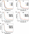
Figure 2. Light absorbance of γ-irradiated serum and γ-irradiated proteins solutions monitored in continuous-scanning mode.
Absorbance of (A) FBS and (B) HS and of the protein solutions containing (C) BSA, (D) BSF, and (E) HSA prepared in either water (solid line) or HEPES buffer (dashed line) was monitored from 200 to 750 nm before and after γ-irradiation at the dose indicated.
Figure 3
Figure 3. Conformational changes of serum proteins following γ-irradiation as revealed by protein fluorometry.
Aliquots of (A) FBS and (B) HS γ-irradiated at the doses indicated were excited with UV light at 280 nm, and the resulting fluorescence emission was monitored between 300 to 450 nm. Control, non-irradiated FBS/HS were included for comparison. Fluorescence was also monitored for both non-irradiated and γ-irradiated solutions of (C) BSA, (D) BSF, and (E) HSA prepared in either water (solid line) or HEPES buffer (dashed line). In most samples, γ-irradiation produced a dose-dependent decrease of fluorescence intensity and a shift of maximum fluorescence to longer wavelengths, consistent with protein unfolding and conformational changes.
Figure 4
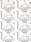
Figure 4. Changes in the secondary structures of serum proteins produced by γ-irradiation as shown by Fourier-transformed infrared spectroscopy.
Aliquots of (A) FBS and (B) HS γ-irradiated at the doses indicated were processed for FTIR analysis. The corresponding, non-irradiated sera were also included for comparison. The analysis was also performed for solutions of (C and D) BSA, (E and F) BSF, and (G and H) HSA, all prepared at 10 mg/ml and γ-irradiated at the doses indicated. The protein solutions were prepared in either (C, E, and G) water or (D, F, and H) HEPES buffer. In order to compare the intensities of the major peak at 3,400 cm−1 seen with the various irradiation doses, the baseline of each spectrum was aligned at 4,000 cm−1. To evaluate the changes seen with the amide-I peak after γ-irradiation, we then normalized the spectra from 1,700 to 1,600 cm−1 using a Gaussian-Lorentzian function (insets) as described in the Materials and Methods . See text for explanation and interpretation.
Figure 5
Figure 5. γ-Irradiation induces the degradation of serum proteins as revealed by SDS-PAGE.
Equal volumes of (A) FBS and (B) HS, either untreated (“0 kGy”) or γ-irradiated at the doses indicated, were electrophoresed on a 10%-polyacrylamide gel in denaturing and reducing conditions. γ-Irradiation produced a dose-dependent degradation of serum proteins as seen through the gradual disappearance of the main protein bands as well as the progressive protein smearing observed in lanes 2 to 7 for both gels. Solutions of HSA, BSA, and BSF, prepared in either (C) water or (D) HEPES buffer were also electrophoresed either before or after γ-irradiation at either 5 or 30 kGy. Note the significant protein profile changes seen as a result of γ-irradiation and the protein solvent used.
Figure 6
Figure 6. γ-Irradiated serum retains its ability to seed NB/NLP in culture.
Aliquots of untreated FBS (“0 kGy”) and γ-FBS (“5-to-50 kGy”) were inoculated into DMEM to concentrations indicated on the top. A650 turbidity readings were performed following inoculation (“Day 1”) or after incubation in cell culture conditions for “1 Month” or “2 Months.” Control, untreated HS and γ-HS were treated in the same way (bottom panel). Notice the time-dependent turbidity increases seen for both non-irradiated and γ-irradiated sera that appear either as bell-shapes or straight lines.
Figure 7
Figure 7. Transient inhibition of NLP formation by γ-irradiated serum.
(A) NLP were prepared from 3 mM each of CaCl2 and NaH2PO4 in DMEM containing either untreated FBS or γ-FBS at the final concentrations indicated on the top. A650 turbidity readings were performed following inoculation (“Day 1”) or after “2 weeks” of incubation in cell culture conditions. (B) Similar experiments were performed with untreated/γ-irradiated HS. Both the untreated sera and the γ-irradiated sera were shown to inhibit NLP formation in a dose-dependent manner (“Day 1”). By 2 weeks, this same inhibitory effect of serum was partially overcome.
Figure 8
Figure 8. Protein profiles of NLP prepared from γ-irradiated serum.
(A) NLP were prepared by adding 3 mM of both CaCl2 and NaH2PO4 into DMEM containing 10% of either untreated FBS (“FBS-NLP”) or γ-FBS (“γ-FBS-NLP”). (B) Particles were prepared in the same manner using either non-irradiated HS (“HS-NLP”) or γ-HS (“γ-HS-NLP”). After incubation, centrifugation and washing, the particles were resuspended into 50 mM EDTA, and processed for SDS-PAGE as described in the Materials and Methods . Note the progressively diffuse protein smears found in the NLP derived from serum that had been irradiated with increasing doses of γ-radiation.
Figure 9
Figure 9. Formation of NLP from γ-irradiated serum proteins in metastable medium versus medium containing submillimolar amounts of calcium and phosphate.
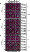
Solutions of (A) BSF, (B) BSA, or (C) both proteins, either untreated or γ-irradiated at the doses indicated on the left, were diluted into DMEM to the concentrations shown on the top. While “None” refers to the absence of additional ion input added to the metastable DMEM, the precipitating reagents CaCl2 and NaH2PO4 were added at either 0.3 or 0.5 mM in some experiments as indicated at the bottom (labeled as “Calcium+Phosphate Added”). A650 turbidity readings were monitored after incubation in cell culture conditions for either one hour (“Day 1”) or “1 Month” as indicated on the right.
Figure 10
Figure 10. Transient inhibition of NLP formation by γ-irradiated serum proteins in supersaturated ionic medium.
(A–C) NLP were prepared from 3 mM each of CaCl2 and NaH2PO4 added into DMEM containing either untreated or γ-irradiated forms of BSF and/or BSA at the final concentrations indicated on the top. The doses of γ-irradiation used are indicated on the left. The 24-well plates were photographed and A650 turbidity readings were performed after incubation in cell culture conditions for 1 hour (“Day 1”) or “3 Days” as indicated. Both non-irradiated (“0 kGy”) and γ-irradiated (“5 and 30 kGy”) proteins inhibited NLP formation in a similar, dose-dependent manner, but this inhibition appeared to have been partially overcome to varying degrees by 3 days of incubation. Note the marked differences in inhibition reversal between (A) BSF and (B) BSA. (C) The results seen with a combination of BSF and BSA appear as summations of the individual effects seen with these proteins.
Figure 11
Figure 11. γ-FBS fails to enhance the formation of NB/NLP cultured from human body fluids.
Body fluids successively filtered through both 0.2 and 0.1-µm membranes were inoculated into DMEM to the concentrations indicated on the top. In some experiments, non-irradiated FBS (“0 kGy”) or γ-FBS (“5-to-50 kGy”) were added into the DMEM solutions to the same concentrations as the indicated body fluids. A650 turbidity readings were performed following inoculation (“Day 1”) or after incubation in cell culture conditions for “2 Months.” Incubation of body fluids, with or without additional FBS/γ-FBS, resulted in similar time-dependent turbidity increases as seen by the straight curves of precipitation seen after 2 months.
Figure 12
Figure 12. NB cultured from either untreated serum or γ-irradiated serum display similar morphologies under electron microscopy.
NB were collected from a 2-month incubation of the indicated serum at 10% as seen in Figure 6. Following centrifugation and washing steps, the NB specimens were processed for (A–F) SEM, (G–L) TEM, or (M–R) thin-section TEM as described in the Materials and Methods . NB obtained from either untreated or γ-irradiated serum appeared as small, rounded and elongated particles displaying varying degrees of crystallinity. Scale bars: 50 nm (J, P); 100 nm (I, K, M–O, R); 200 nm (A–E, G, H, L, Q); 500 nm (F).
Figure 13
Figure 13. NB cultured from either untreated serum or γ-irradiated serum show similar elemental compositions when analyzed by energy-dispersive X-ray spectroscopy.
NB were retrieved from the experiments described in Figure 6. Precipitates were taken from 2-month-old cultures of either untreated FBS (A) or from FBS γ-irradiated at either 30 kGy (B) or 50 kGy (C). In (D–F), NB specimens were obtained from cultures of untreated HS (D) or from HS γ-irradiated at either 30 kGy (E) or 50 kGy (F). Following centrifugation and washing, the specimens were processed for EDX as described in the Materials and Methods . Major peaks of carbon (C), oxygen (O), phosphorus (P), and calcium (Ca) were noticed for the various NB specimens, consistent with the presence of a calcium phosphate mineral containing carbonate ions. The following Ca/P ratios were obtained: (A) 1.40; (B) 1.40; (C) 1.24; (D) 0.98; (E) 0.86; and (F) 1.09.

References

    1. Martel J, Young JD. Purported nanobacteria in human blood as calcium carbonate nanoparticles. Proc Natl Acad Sci U S A. 2008;105:5549–5554. - PMC - PubMed
    1. Young JD, Martel J, Young L, Wu CY, Young A, et al. Putative nanobacteria represent physiological remnants and culture by-products of normal calcium homeostasis. PLoS ONE. 2009;4:e4417. - PMC - PubMed
    1. Young JD, Martel J, Young D, Young A, Hung CM, et al. Characterization of granulations of calcium and apatite in serum as pleomorphic mineralo-protein complexes and as precursors of putative nanobacteria. PLoS ONE. 2009;4:e5421. - PMC - PubMed
    1. Wu CY, Martel J, Young D, Young JD. Fetuin-A/albumin-mineral complexes resembling serum calcium granules and putative nanobacteria: demonstration of a dual inhibition-seeding concept. PLoS ONE. 2009;4:e8058. - PMC - PubMed
    1. Young JD, Martel J. The rise and fall of nanobacteria. Sci Am. 2010;302:52–59. - PubMed

Publication types