. 2010 Apr 26;5(4):e10347.
doi: 10.1371/journal.pone.0010347.

Isoform-specific upregulation of palladin in human and murine pancreas tumors

Affiliations

Isoform-specific upregulation of palladin in human and murine pancreas tumors

Silvia M Goicoechea et al. PLoS One. .

Abstract

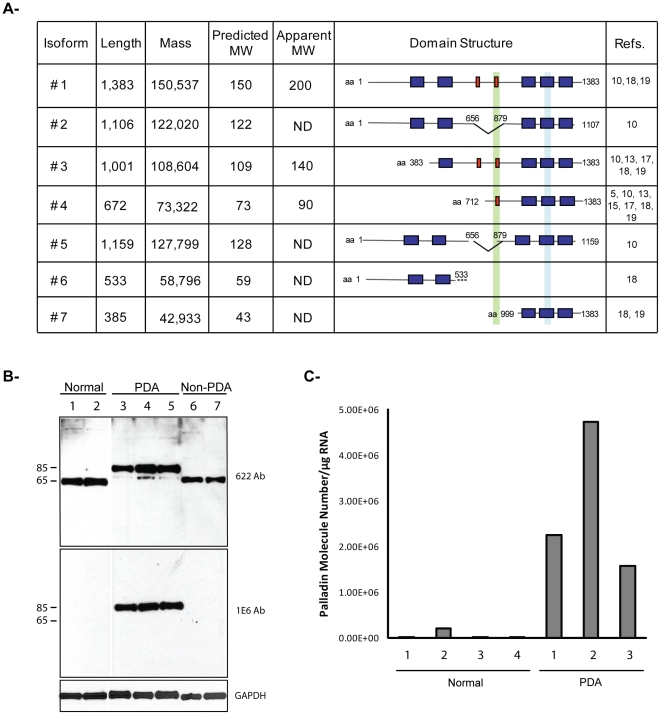
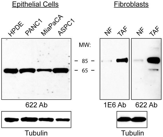
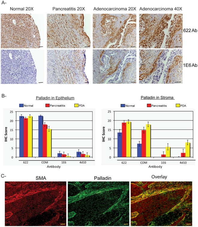
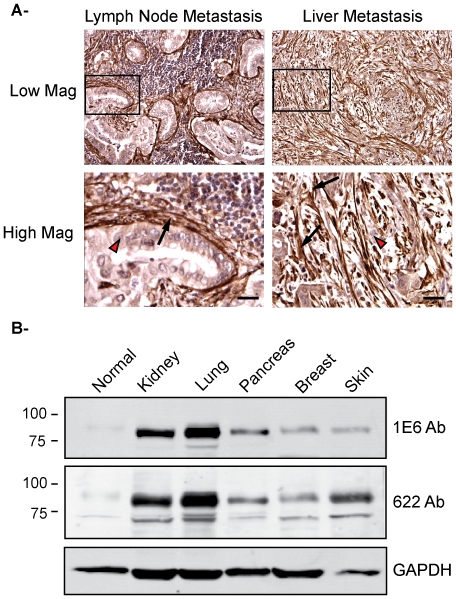
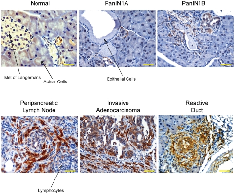
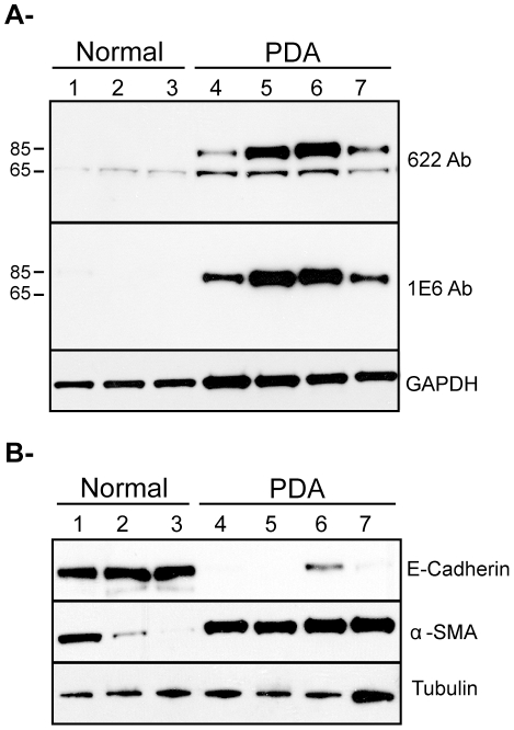
Pancreatic ductal adenocarcinoma (PDA) is a lethal disease with a characteristic pattern of early metastasis, which is driving a search for biomarkers that can be used to detect the cancer at an early stage. Recently, the actin-associated protein palladin was identified as a candidate biomarker when it was shown that palladin is mutated in a rare inherited form of PDA, and overexpressed in many sporadic pancreas tumors and premalignant precursors. In this study, we analyzed the expression of palladin isoforms in murine and human PDA and explored palladin's potential use in diagnosing PDA. We performed immunohistochemistry and immunoblot analyses on patient samples and tumor-derived cells using an isoform-selective monoclonal antibody and a pan-palladin polyclonal antibody. Immunoblot and real-time quantitative reverse transcription-PCR were used to quantify palladin mRNA levels in human samples. We show that there are two major palladin isoforms expressed in pancreas: 65 and 85-90 kDa. The 65 kDa isoform is expressed in both normal and neoplastic ductal epithelial cells. The 85-90 kDa palladin isoform is highly overexpressed in tumor-associated fibroblasts (TAFs) in both primary and metastatic tumors compared to normal pancreas, in samples obtained from either human patients or genetically engineered mice. In tumor-derived cultured cells, expression of palladin isoforms follows cell-type specific patterns, with the 85-90 kDa isoform in TAFs, and the 65 kDa isoform predominating in normal and neoplastic epithelial cells. These results suggest that upregulation of 85-90 kDa palladin isoform may play a role in the establishment of the TAF phenotype, and thus in the formation of a desmoplastic tumor microenvironment. Thus, palladin may have a potential use in the early diagnosis of PDA and may have much broader significance in understanding metastatic behavior.


Conflict of interest statement

Competing Interests: The authors have declared that no competing interests exist.

Figures

Figure 1
Figure 1. Analysis of human palladin isoforms in pancreatic tissues.
A. Human palladin isoforms. Proline-rich domains are represented by red boxes, and Ig-like domains are shown as blue boxes. The epitope recognized by the 1E6 and 4D10 antibodies is highlighted in green. The region amplified by RT-qPCR is highlighted in light blue. Isoform #1, 3 and 4 are the primary products of the palladin gene and have been detected by immunoblotting. The sequences of these isoforms are published. The sequences of isoforms #2, 5, 6, and 7 were obtained from genomic databases. “ND”: not-determined. B. Western blot analysis of pancreas samples. Small pieces of fresh tissue were snap-frozen in liquid nitrogen, ground in a chilled mortar and pestle, extracted in a detergent-containing lysis buffer, and centrifuged at 15,000×g to remove any unsolubilized particulates. The supernatant was boiled in Laemmli sample buffer and resolved by SDS-PAGE, with 15 µg protein loaded per lane. The samples were immunoblotted and probed with two anti-palladin antibodies and an antibody to GADPH (a housekeeping gene) as a control for equal loading. Lanes 1–2: normal pancreas. Lanes 3–5: primary adenocarcinoma tumors (PDA). Lane 6–7: Non-primary adenocarcinoma tumors (Non-PDA) (Lane 6: solid pseudopapillary tumor, Lane 7: neuroendocrine tumor). C. RT-qPCR. Total RNA was isolated from normal tissue (patients 1–4) and PDA tumors (patients 1–3), reverse transcribed, and subjected to RT-qPCR using gene-specific primers. Each bar represents the mean + SEM (0.06–0.35%) from three or more independent determinations.
Figure 2
Figure 2. Palladin expression in pancreatic cancer cells.
Immunoblot analysis of pancreatic tumor-derived cell lines, tumor-associated fibroblasts, and corresponding normal cells. Pancreatic cells: normal human pancreatic ductal epithelial cells (HPDE) and three tumor cell lines: PANC1, MiaPaCA, and ASPC1. Fibroblasts: normal adult fibroblasts (NF) and tumor-associated fibroblasts (TAF). Whole cell lysates were analyzed by western blot using the polyclonal antibody 622. Blots were also stained for tubulin as a control for equal loading.
Figure 3
Figure 3. Palladin staining of paraffin-embedded patient tissues.
A. IHC staining was performed using standard antigen-retrieval protocols, and counter-stained with hematoxylin. Tissue sections were stained for palladin using two palladin antibodies: polyclonal 622 and monoclonal 1E6. Palladin stain is detected with brown reaction product. In tumor sections, palladin is detected at dramatically elevated levels in the stromal fibroblasts. Note also the expanded stroma around the neoplastic cells, which is characteristic of the desmoplastic reaction. Scale bars, 200 µM. B. Quantification of immunohistochemistry results. Ten sections each of normal pancreatitis and adenocarcinoma specimens were stained with four different antibodies (622, COM, 1E6 and 4D10) and scored by two pathologists, as described in the text. Results for both ductal epithelium (left) and stroma (right) stained with various palladin antibodies are shown for normal pancreas (n = 9, blue), pancreatitis (n = 7, red) and pancreatic adenocarcinoma (n = 10, yellow). The results confirmed that palladin levels are increased in the stroma, and not the epithelial tumor cells, of the adenocarcinomas. Although palladin levels are also increased in cases of chronic pancreatitis, they do not reach the same levels as in the tumors. Compared to the polyclonal 622 and COM, the monoclonal antibodies 1E6 and 4D10 are effective at distinguishing between pancreatitis and cancer. C. Double-label immunostaining for palladin (1E6 Ab) and α-SMA in sections of pancreatic tumors confirms that palladin is strongly detected in a population of activated TAFs that surround the neoplastic cells. Scale bars, 200 µM.
Figure 4
Figure 4. Palladin in human pancreatic cancer metastasis and other human cancers.
A. Immunohistochemistry of lymph node and liver metastases. Formalin-fixed, paraffin-embedded tissue sections were stained with COM antibody. Left: Low and high magnification images of lymph node metastasis. Right: Low and high magnification of liver metastasis. Arrow heads, lightly-stained tumor epithelial cells. Arrows, intensely-stained tumor-associated fibroblasts. Scale bars, 100 µM. B. Immunoblot analysis of tumor-associated fibroblasts from different human cancers. Tumor-associated fibroblasts were isolated from different human cancers: normal, kidney, lung, pancreas, breast and skin. Whole cell lysates were analyzed by western blot using the monoclonal antibody 1E6 and the polyclonal antibody 622. Blots were also stained for GAPDH as a control for equal loading.
Figure 5
Figure 5. Palladin expression in LSL-KrasG12D/+ mice.
Normal, primary tumors and metastasis tissue sections were stained with 622 Ab. Palladin staining is noted in stromal fibroblasts in both primary adenocarcinomas and metastases. Scale bars, 50 µM.
Figure 6
Figure 6. Detection of palladin in post-surgical samples collected with 18-gauge needles.
A. Samples of normal (lane 1 to 3) and pancreatic adenocarcinoma (lanes 4 to 7) were obtained from donated post-surgical organs using 18-gauge needles. Tissue samples were snap-frozen, ground, lysed and analyzed as in Figure 1. The blot was stained with both monoclonal (1E6) and polyclonal (622) palladin antibodies, and the major band (85–90 kDa) was detected by both antibodies in all tumor samples. B. Same samples (normal, lane 1–3 and PDA, lane 4–7) were analyzed for epithelial vs myofibroblast markers. The blot was stained with both, anti-E-cadherin antibody (as an epithelial cell marker) and anti-αSMA antibody (as a myofibroblast marker). Blots were stained for tubulin as a control for equal loading.

References

    1. Jemal A, Siegel R, Ward E, Hao Y, Xu J, et al. Cancer statistics, 2009. CA Cancer J Clin. 2009;59:225–49. - PubMed
    1. Cleary SP, Gryfe R, Guindi M, Greig P, Smith L, et al. Prognostic factors in resected pancreatic adenocarcinoma: analysis of actual 5-year survivors. J Am Coll Surg. 2004;198:722–731. - PubMed
    1. Conlon KC, Klimstra DS, Brennan MF. Long-term survival after curative resection for pancreatic ductal adenocarcinoma. Clinicopathologic analysis of 5-year survivors. Ann Surg. 1996;223:273–279. - PMC - PubMed
    1. Sebolt-Leopold JS, English JM. Mechanisms of drug inhibition of signalling molecules. Nature. 2006;441:457–462. - PubMed
    1. Pogue-Geile KL, Chen R, Bronner MP, Crnogorac-Jurcevic T, Moyes KW, et al. Palladin mutation causes familial pancreatic cancer and suggests a new cancer mechanism. PLoS Med. 2006;3:e516. - PMC - PubMed

Publication types