. 2010 Apr 26;5(4):e10358.
doi: 10.1371/journal.pone.0010358.

Genetic analysis of the early natural history of epithelial ovarian carcinoma

Affiliations

Genetic analysis of the early natural history of epithelial ovarian carcinoma

Bhavana Pothuri et al. PLoS One. .

Abstract

Background: The high mortality rate associated with epithelial ovarian carcinoma (EOC) reflects diagnosis commonly at an advanced stage, but improved early detection is hindered by uncertainty as to the histologic origin and early natural history of this malignancy.

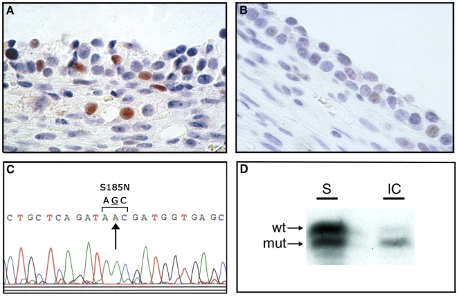
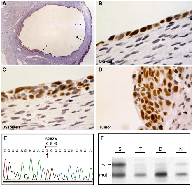
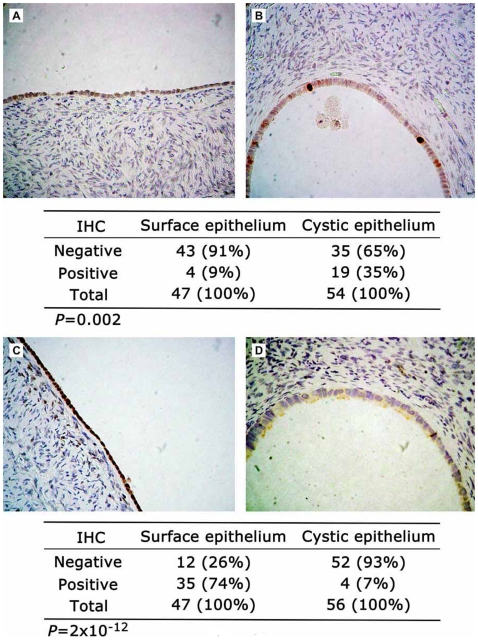
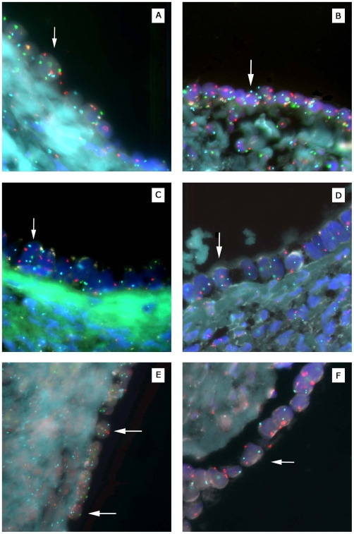
Methodology/principal findings: Here we report combined molecular genetic and morphologic analyses of normal human ovarian tissues and early stage cancers, from both BRCA mutation carriers and the general population, indicating that EOCs frequently arise from dysplastic precursor lesions within epithelial inclusion cysts. In pathologically normal ovaries, molecular evidence of oncogenic stress was observed specifically within epithelial inclusion cysts. To further explore potential very early events in ovarian tumorigenesis, ovarian tissues from women not known to be at high risk for ovarian cancer were subjected to laser catapult microdissection and gene expression profiling. These studies revealed a quasi-neoplastic expression signature in benign ovarian cystic inclusion epithelium compared to surface epithelium, specifically with respect to genes affecting signal transduction, cell cycle control, and mitotic spindle formation. Consistent with this gene expression profile, a significantly higher cell proliferation index (increased cell proliferation and decreased apoptosis) was observed in histopathologically normal ovarian cystic compared to surface epithelium. Furthermore, aneuploidy was frequently identified in normal ovarian cystic epithelium but not in surface epithelium.

Conclusions/significance: Together, these data indicate that EOC frequently arises in ovarian cystic inclusions, is preceded by an identifiable dysplastic precursor lesion, and that increased cell proliferation, decreased apoptosis, and aneuploidy are likely to represent very early aberrations in ovarian tumorigenesis.


Conflict of interest statement

Competing Interests: The authors have declared that no competing interests exist.

Figures

Figure 1
Figure 1. Assessment of p53/TP53 status and BRCA2 allelotype in epithelial inclusion cysts of normal ovary from a BRCA2 6174delT heterozygote (specimen PO67 in Supplemental Table S1).
(A) p53 immunopositive cells from an epithelial inclusion cyst. (B) p53 immunonegative cells from a different epithelial inclusion cyst from the same ovary. (C) Sequence analysis of TP53 in p53 immunopositive cells from inclusion cyst shown in panel (A). A missense mutation is evident at codon 185 (AGC→AAC; S185N). (D) Loss of the wild-type (wt) BRCA2 allele in DNA derived from the inclusion cyst (IC) shown in panel (A), with retention of mutant (mut) and wild-type alleles in DNA derived from surface (S) epithelium from the same ovary.
Figure 2
Figure 2. Morphological, immunohistochemical, and genetic analyses of early stage ovarian carcinoma arising in a BRCA1 185delAG heterozygote (specimen OC16 in Supplementary Table S2).
(A) Low-power photomicrograph of histologic progression of normal epithelium (N) to dysplastic epithelium (D) to invasive carcinoma (T) arising within an inclusion cyst. (B,C,D) High-power photpmicrographs of cellular regions of normal, dysplastic, and carcinoma, respectively, as shown in panel (A). The immunostain is for p53 in panels (A–D). (E) Sequence analysis of TP53 representative of DNA samples from all three cellular components shown in panels (B–D). A missense mutation is evident at codon 282 (CGG→TGG; R282W). (F) Loss of the wild-type (wt) BRCA1 allele in DNA derived from normal, dysplastic, and tumor cells shown in panels (B–D), with retention of mutant (mut) and wild-type alleles in surface epithelium from the same ovary.
Figure 3
Figure 3. Semiquantitative immunohistochemical analyses of gene products differentially expressed in normal ovarian cystic epithelium and ovarian carcinoma compared to normal ovarian surface epithelium.
(A) Normal ovarian surface epithelium displaying negative immunstaining for TOP2A. (B) Normal ovarian cystic epithelium displaying strong immunopositivity for TOP2A in two cells. (C) Normal ovarian surface epithelium displaying immunopositivity for FOS in all cells. (D) Normal ovarian cystic epithelium displaying negative immunostaining for FOS. Indicated under each pair of photomicrographs are the proportions of immunopositive and immunonegative cellular compartments for the indicated number of ovarian specimens, with the associated P-values.
Figure 4
Figure 4. Fluorescence in situ hybridization analysis of ploidy in normal ovarian cystic and surface epithelia.
All examples shown are from distinct ovarian specimens. (A) Cystic epithelial cell (arrow) from sample #112 (see Table 2) with three copies of chromosomes 3 (red) and 11 (green). (B) Cystic epithelial cell from sample #84 with three copies of chromosomes 6 (blue) and 11 (green). (C) Cystic epithelial cell from sample #135 with three copies of chromosome 6 (blue). (D) Cystic epithelial cell from sample #59 with three copies of chromosome 8 (blue). (E) Surface epithelial cells from sample # 13 with two copies of chromosomes 3 (red), 6 (blue), and 11 (green). (F) Surface epithelial cell from sample #102 with two copies of chromosomes 3 (red) and 8 (blue).

References

    1. Fleming GF, Ronnett BM, Seidman J, Zaino R, Rubin SC. Epithelial ovarian cancer. In: Barakat RR, Markman M, Randall ME, editors. Principles and practice of gynecologic oncology, 5th ed. Baltimore: Lippincott, Williams & Wilkins; 2009. pp. 763–835.
    1. Rosenthal AN, Menon U, Jacobs IJ. Screening for ovarian cancer. Clin Obstet Gynecol. 2006;49:433–447. - PubMed
    1. Kohn EC, Azad N, Annunziata C, Dhamoon AS, Whiteley G. Proteomics as a tool for biomarker discovery. Dis Markers. 2007;23:411–417. - PMC - PubMed
    1. Visintin I, Feng Z, Longton G, Ward DC, Alvero AB, et al. Diagnostic markers for early detection of ovarian cancer. Clin Cancer Res. 2008;14:1065–1072. - PubMed
    1. Kurman RJ, Shih Ie-M. The origin and pathogenesis of epithelial ovarian cancer: a proposed unifying theory. Am J Surg Pathol. 2010;34:433–443. - PMC - PubMed

Publication types