

Ultrasonography: a novel approach to central venous cannulation
- PMID: 20436690
- PMCID: PMC2856149
- DOI: 10.4103/0972-5229.60174
Ultrasonography: a novel approach to central venous cannulation
Abstract
Background: Portable ultrasound machines are highly valuable in ICUs, where a patient's condition might not permit shifting the patient to the USG department for imaging. Traditionally central lines are put blindly using anatomical landmarks, which often result in complications such as difficulty in access, misplaced lines, pneumothorax, bleeding from inadvertent arterial punctures, etc. Ultrasonography provides "real time" imaging, i.e., the needle can be visualized entering the vein.
Aims: We performed a study to compare USG guided central venous cannulation (CVC) and conventional anatomical landmark approach to CVC, in terms of ease of cannulation, time consumed, and associated complications.
Settings and design: The study was performed in a 16-bed open ICU. Eighty patients were randomly divided in two groups.
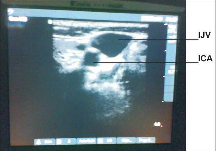
Materials and methods: The right internal jugular vein (IJV) was cannulated in all. In Group I, a portable ultrasound machine was used during cannulation. The vessels were visualized in the transverse section with the internal carotid artery (ICA) identified as a circular pulsatile structure, while the IJV as a lateral, oval nonpulsatile structure). The needle was inserted perpendicular to the skin under visualization on the US screen. Central venous line was then inserted by the Seldinger technique. In Group II, CVC was performed by the conventional landmark approach. The parameters studied included time for insertion, attempts required, and complications encountered.
Statistical analysis: The database of all parameters was analyzed using SPSS software version 10.5.
Results: The mean time to successful insertion was 145 and 176.4 sec in groups I and II, respectively (p = 0.00). An average of 1.2 attempts per cannulation was required for group I, while 1.53 for group II (p = 0.03): 10% witnessed arterial puncture and 2.5% pneumothorax in group I and none in group II.
Conclusion: USG-guided CVC is thus easier, quicker, and safer than landmark approach.
Keywords: central venous cannulation; intensive care unit; ultrasound.
Conflict of interest statement
Figures

References
-
- Milling TJ, Jr, Rose J, Briggs WM, Birkhahn R, Gaeta TJ, Bove JJ, et al. Randomized, controlled clinical trial of point-of-care limited ultrasonography assistance of central venous cannulation: The third sonography outcomes assessment program (SOAP-3) trial. Crit Care Med. 2005;33:1764–9. - PubMed
-
- Julie L, Martin D, Andrew F. Real time ultrasonographically guided internal jugular vein catheterization in the emergency department increases success rates and reduces complications: A randomized, prospective study. Ann Emerg Med. 2006;48:540–7. - PubMed
-
- Gilbert TB, Seneff MG, Becker RB. Facilitation of internal jugular venous cannulation using an audio-guided doppler ultrasound vascular access device: Results from a prospective, dual-center, randomized, crossover clinical study. Crit Care Med. 1995;23:60–5. - PubMed
-
- Jefferson P, Ogbue MN, Hamilton KE, Ball DR. A survey of the use of portable ultrasound for central vein cannulation on critical care units in the UK. Anaesthesia. 2002;57:365–8. - PubMed
LinkOut - more resources
Full Text Sources
Miscellaneous